• 文献检索
  • 文档翻译
  • 深度研究
  • 学术资讯
  • Suppr Zotero 插件Zotero 插件
  • 邀请有礼
  • 套餐&价格
  • 历史记录
应用&插件
Suppr Zotero 插件Zotero 插件浏览器插件Mac 客户端Windows 客户端微信小程序
定价
高级版会员购买积分包购买API积分包
服务
文献检索文档翻译深度研究API 文档MCP 服务
关于我们
关于 Suppr公司介绍联系我们用户协议隐私条款
关注我们

Suppr 超能文献

核心技术专利:CN118964589B侵权必究
粤ICP备2023148730 号-1Suppr @ 2026

文献检索

告别复杂PubMed语法,用中文像聊天一样搜索,搜遍4000万医学文献。AI智能推荐,让科研检索更轻松。

立即免费搜索

文件翻译

保留排版,准确专业,支持PDF/Word/PPT等文件格式,支持 12+语言互译。

免费翻译文档

深度研究

AI帮你快速写综述,25分钟生成高质量综述,智能提取关键信息,辅助科研写作。

立即免费体验

负重磁共振成像作为一种在脊柱解剖结构中产生生物力学变化的诊断工具。

Weight-Bearing Magnetic Resonance Imaging as a Diagnostic Tool That Generates Biomechanical Changes in Spine Anatomy.

作者信息

Fiani Brian, Griepp Daniel W, Lee Jason, Davati Cyrus, Moawad Christina M, Kondilis Athanasios

机构信息

Neurosurgery, Desert Regional Medical Center, Palm Springs, USA.

Neurosurgery, College of Osteopathic Medicine, New York Institute of Technology, Old Westbury, USA.

出版信息

Cureus. 2020 Dec 14;12(12):e12070. doi: 10.7759/cureus.12070.

DOI:10.7759/cureus.12070
PMID:33489488
原文链接:https://pmc.ncbi.nlm.nih.gov/articles/PMC7805418/
Abstract

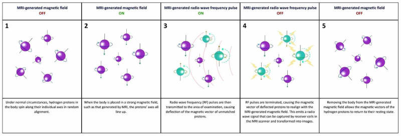
Weight-bearing magnetic resonance imaging (MRI) is a unique modality in diagnostic imaging that allows for the assessment of spinal pathology in ways considered previously inaccessible or insufficient with the conventional MRI technique. Due to limitations in positioning within the MRI machine itself, difficulties would be posed in determining the underlying cause of a patient's pain or neurological symptoms, as the traditional supine position utilized can, in many cases, alleviate the severity of presented symptoms. Weight-bearing MRI addresses this concern by allowing a clinician to position a patient (to a certain degree) into flexion, extension, rotation, or side-bending with an axial load that can mimic physiologic conditions in order to replicate the conditions the patient experiences in order to give clinicians a clearer understanding of the anatomical relationship of the spine and surrounding tissues that may lead to a particular presentation of symptoms. These findings can then guide treatment approaches that are better tailored to a patient's needs in order to initiate treatment earlier and shorten the duration of treatment necessary for patient benefit. The goal of this review is to describe and differentiate weight-bearing MRI from conventional MRI as well as examine the advantages and disadvantages of either imaging modality. This will include assessing cost-effectiveness and improvements in clinical outcomes. Further, the advancements of weight-bearing MRI will be discussed, including potentially unique clinical applications in development.

摘要

负重磁共振成像(MRI)是诊断成像中的一种独特方式,它能够以传统MRI技术以前被认为无法实现或不够充分的方式评估脊柱病变。由于MRI机器本身在定位方面存在局限性,确定患者疼痛或神经症状的潜在原因会面临困难,因为在许多情况下,所采用的传统仰卧位能够减轻所呈现症状的严重程度。负重MRI通过允许临床医生在轴向负荷下将患者(在一定程度上)置于屈曲、伸展、旋转或侧弯姿势来解决这一问题,这种轴向负荷可以模拟生理状况,以便复制患者所经历的情况,从而使临床医生更清楚地了解可能导致特定症状表现的脊柱与周围组织的解剖关系。这些发现随后可以指导更符合患者需求的治疗方法,以便更早地开始治疗并缩短为使患者受益所需的治疗时间。本综述的目的是描述负重MRI并将其与传统MRI区分开来,同时研究这两种成像方式的优缺点。这将包括评估成本效益和临床结果的改善情况。此外,还将讨论负重MRI的进展,包括正在开发中的潜在独特临床应用。

https://cdn.ncbi.nlm.nih.gov/pmc/blobs/9744/7805418/65368706606e/cureus-0012-00000012070-i01.jpg

相似文献

1
Weight-Bearing Magnetic Resonance Imaging as a Diagnostic Tool That Generates Biomechanical Changes in Spine Anatomy.负重磁共振成像作为一种在脊柱解剖结构中产生生物力学变化的诊断工具。
Cureus. 2020 Dec 14;12(12):e12070. doi: 10.7759/cureus.12070.
2
Weight-bearing MRI of the Lumbar Spine: Technical Aspects.腰椎负重磁共振成像:技术要点
Semin Musculoskelet Radiol. 2019 Dec;23(6):609-620. doi: 10.1055/s-0039-1697936. Epub 2019 Nov 19.
3
Proceedings of the State-of-the-Art Symposium on Diagnostic and Interventional Radiology of the Spine, Antwerp, September 7, 2002 (Part two). Upright, weight-bearing, dynamic-kinetic MRI of the spine: pMRI/kMRI.《脊柱诊断与介入放射学最新技术研讨会会议记录》,安特卫普,2002年9月7日(第二部分)。脊柱直立位、负重、动态-动力磁共振成像:pMRI/kMRI。
JBR-BTR. 2003 Sep-Oct;86(5):286-93.
4
Magnetic resonance imaging of the weight-bearing spine.负重脊柱的磁共振成像。
Semin Musculoskelet Radiol. 2003 Dec;7(4):277-86. doi: 10.1055/s-2004-815675.
5
Weight-bearing MRI of the Lumbar Spine: Spinal Stenosis and Spondylolisthesis.腰椎负重磁共振成像:椎管狭窄与椎体滑脱
Semin Musculoskelet Radiol. 2019 Dec;23(6):621-633. doi: 10.1055/s-0039-1697937. Epub 2019 Nov 19.
6
The effect of body position and axial load on spinal canal morphology: an MRI study of central spinal stenosis.体位和轴向负荷对椎管形态的影响:一项关于中央型椎管狭窄的MRI研究
Spine (Phila Pa 1976). 2008 Jan 1;33(1):61-7. doi: 10.1097/BRS.0b013e31815e395f.
7
Dynamic change of dural sac cross-sectional area in axial loaded magnetic resonance imaging correlates with the severity of clinical symptoms in patients with lumbar spinal canal stenosis.轴向加载磁共振成像中硬脊膜囊横截面积的动态变化与腰椎椎管狭窄症患者临床症状的严重程度相关。
Spine (Phila Pa 1976). 2012 Feb 1;37(3):207-13. doi: 10.1097/BRS.0b013e3182134e73.
8
Dynamic Cervical Cord Compression Post-laminectomy Visualized by Flexion-extension Magnetic Resonance Imaging: Case Report.屈伸位磁共振成像显示的椎板切除术后动态颈髓压迫:病例报告
Cureus. 2019 Jan 14;11(1):e3878. doi: 10.7759/cureus.3878.
9
Physiologic imaging of the spine.脊柱的生理成像
Radiol Clin North Am. 2012 Jul;50(4):599-611. doi: 10.1016/j.rcl.2012.04.004.
10
Axial loading during magnetic resonance imaging in patients with lumbar spinal canal stenosis: does it reproduce the positional change of the dural sac detected by upright myelography?轴向加载在腰椎管狭窄症患者磁共振成像中的应用:它是否能重现直立性椎管造影检测到的硬脊膜囊位置变化?
Spine (Phila Pa 1976). 2012 Jul 15;37(16):E985-92. doi: 10.1097/BRS.0b013e31821038f2.

引用本文的文献

1
Optimizing infant neuroimaging methods to understand the neurodevelopmental impacts of early nutrition and feeding.优化婴儿神经成像方法以了解早期营养和喂养对神经发育的影响。
Dev Cogn Neurosci. 2025 Jan;71:101481. doi: 10.1016/j.dcn.2024.101481. Epub 2024 Nov 23.
2
Seeing the unseen: The role of bioimaging techniques for the diagnostic interventions in intervertebral disc degeneration.洞察不可见:生物成像技术在椎间盘退变诊断干预中的作用。
Bone Rep. 2024 Jun 27;22:101784. doi: 10.1016/j.bonr.2024.101784. eCollection 2024 Sep.
3
Effects of axial loading and positions on lumbar spinal stenosis: an MRI study using a new axial loading device.

本文引用的文献

1
Cost Effectiveness of Mobile versus Fixed Computed Tomography and Magnetic Resonance Imaging: A Systematic Review.移动与固定计算机断层扫描及磁共振成像的成本效益:系统评价
Iran J Public Health. 2019 Aug;48(8):1418-1427.
2
Weight-bearing MRI of the Lumbar Spine: Technical Aspects.腰椎负重磁共振成像:技术要点
Semin Musculoskelet Radiol. 2019 Dec;23(6):609-620. doi: 10.1055/s-0039-1697936. Epub 2019 Nov 19.
3
Diagnostic Importance of Axial Loaded Magnetic Resonance Imaging in Patients with Suspected Lumbar Spinal Canal Stenosis.
轴向负荷及体位对腰椎管狭窄症的影响:一项使用新型轴向负荷装置的磁共振成像研究
Skeletal Radiol. 2025 Feb;54(2):199-208. doi: 10.1007/s00256-024-04720-5. Epub 2024 Jun 7.
4
Diagnostic value of a new axial loading MRI device in patients with suspected lumbar spinal stenosis.新型轴向加载 MRI 设备在疑似腰椎椎管狭窄症患者中的诊断价值。
Eur Radiol. 2023 May;33(5):3200-3210. doi: 10.1007/s00330-023-09447-w. Epub 2023 Feb 23.
5
Axial loading lumbar magnetic resonance imaging with a new device in asymptomatic individuals.使用一种新设备对无症状个体进行轴向负荷腰椎磁共振成像。
Quant Imaging Med Surg. 2023 Jan 1;13(1):58-65. doi: 10.21037/qims-22-283. Epub 2022 Sep 22.
6
Can Swallowing Cerebral Neurophysiology Be Evaluated during Ecological Food Intake Conditions? A Systematic Literature Review.能否在生态食物摄入条件下评估吞咽脑神经病生理学?一项系统文献综述。
J Clin Med. 2022 Sep 18;11(18):5480. doi: 10.3390/jcm11185480.
轴向加载磁共振成像对疑似腰椎管狭窄症患者的诊断意义。
World Neurosurg. 2019 Jul;127:e69-e75. doi: 10.1016/j.wneu.2019.02.091. Epub 2019 Mar 9.
4
Successful visualization of dynamic change of lumbar nerve root compression with the patient in both upright and prone positions using dynamic digital tomosynthesis-radiculography in patients with lumbar foraminal stenosis: An initial report of three cases.在腰椎管狭窄症患者中使用动态数字断层合成神经根造影术在患者直立和俯卧位时成功可视化腰神经根压迫的动态变化:三例初步报告
J Clin Neurosci. 2019 Apr;62:256-259. doi: 10.1016/j.jocn.2018.12.016. Epub 2019 Jan 9.
5
Preoperative Assessment of Neural Elements in Lumbar Spinal Stenosis by Upright Magnetic Resonance Imaging: An Implication for Routine Practice?直立位磁共振成像对腰椎管狭窄症神经结构的术前评估:对常规实践有何启示?
Cureus. 2018 Apr 6;10(4):e2440. doi: 10.7759/cureus.2440.
6
Introducing Standing Weight-bearing MRI in the Di-agnostics of Low Back Pain and Degenerative Spinal Disorders.站立位负重磁共振成像在腰痛和退行性脊柱疾病诊断中的应用介绍。
Dan Med J. 2017 Oct;64(10).
7
An increase in the degree of olisthesis during axial loading reduces the dural sac size and worsens clinical symptoms in patients with degenerative spondylolisthesis.轴向加载过程中olisthesis 程度的增加会减小硬脊膜囊的大小,并使退行性脊椎滑脱症患者的临床症状恶化。
Spine J. 2018 May;18(5):726-733. doi: 10.1016/j.spinee.2017.08.251. Epub 2017 Sep 21.
8
Changes in dural sac caliber with standing MRI improve correlation with symptoms of lumbar spinal stenosis.站立位MRI时硬脊膜囊管径的变化可改善与腰椎管狭窄症状的相关性。
Eur Spine J. 2017 Oct;26(10):2666-2675. doi: 10.1007/s00586-017-5211-7. Epub 2017 Jul 12.
9
Conventional Supine MRI With a Lumbar Pillow-An Alternative to Weight-bearing MRI for Diagnosing Spinal Stenosis?: A Cross-sectional Study.使用腰枕的传统仰卧位磁共振成像:诊断腰椎管狭窄症时负重磁共振成像的替代方法?一项横断面研究。
Spine (Phila Pa 1976). 2017 May 1;42(9):662-669. doi: 10.1097/BRS.0000000000001889.
10
Upright magnetic resonance imaging of the lumbar spine: Back pain and radiculopathy.腰椎的直立位磁共振成像:背痛与神经根病
J Craniovertebr Junction Spine. 2016 Jan-Mar;7(1):31-7. doi: 10.4103/0974-8237.176619.