Dos Santos Pereira Maurício, Abreu Gabriel Henrique Dias, Rocca Jeremy, Hamadat Sabah, Raisman-Vozari Rita, Michel Patrick Pierre, Del Bel Elaine
Department of Basic and Oral Biology, FORP, Campus USP, University of São Paulo, Ribeirão Preto, Brazil.
Department of Physiology, FMRP, Campus USP, University of São Paulo, Ribeirão Preto, Brazil.
Front Pharmacol. 2021 Jan 11;11:617085. doi: 10.3389/fphar.2020.617085. eCollection 2020.
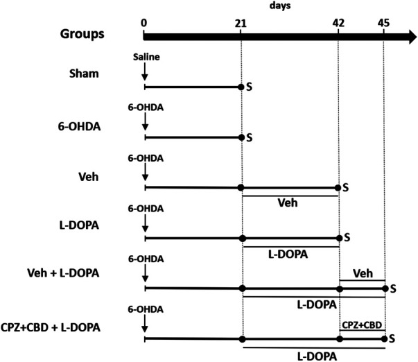
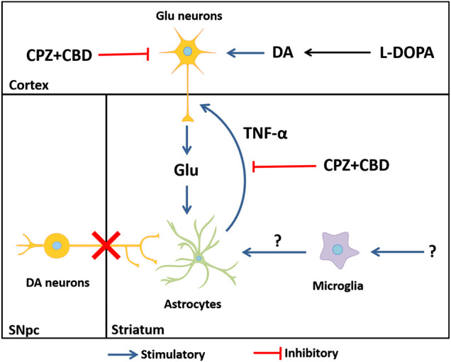
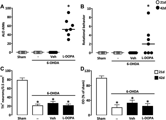
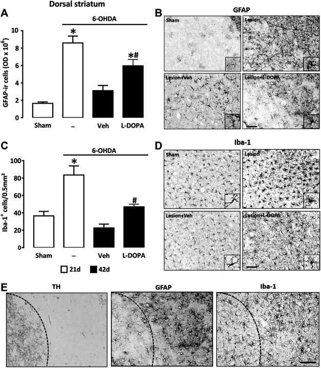
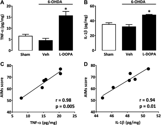
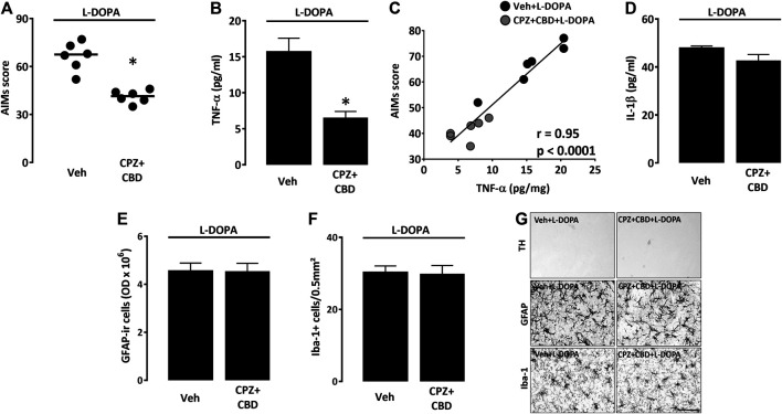
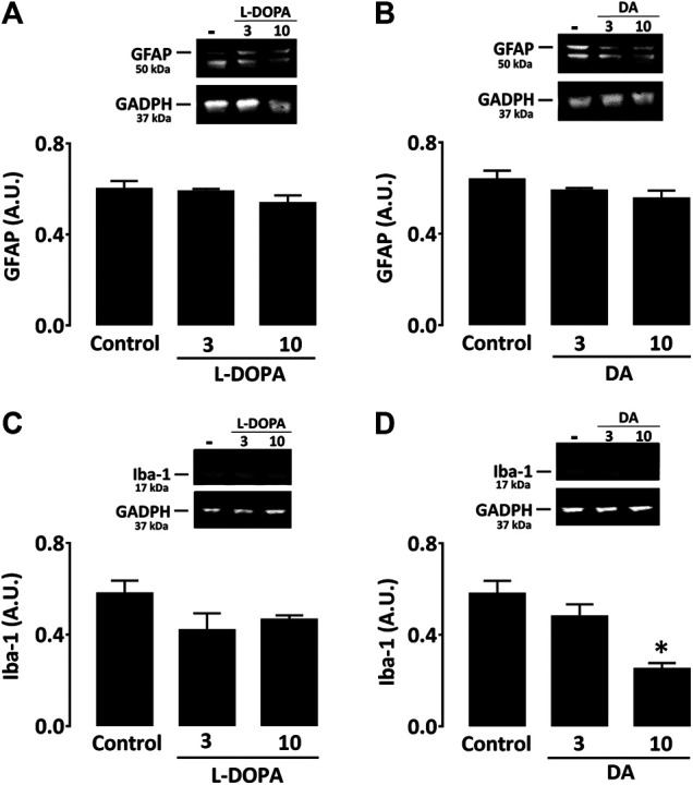
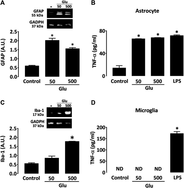
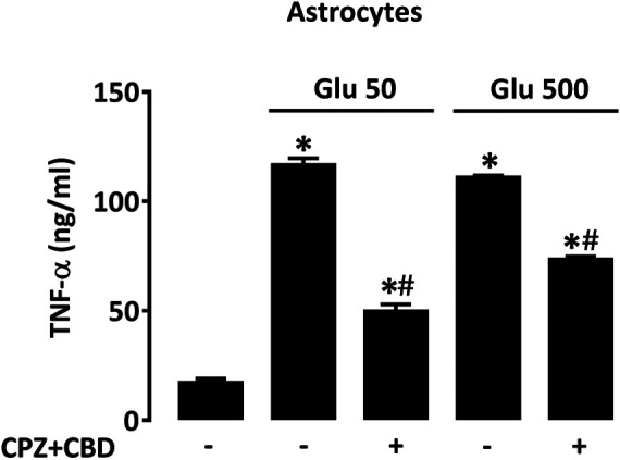
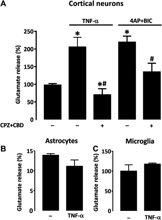
Our present objective was to better characterize the mechanisms that regulate striatal neuroinflammation in mice developing L-DOPA-induced dyskinesia (LID). For that, we used 6-hydroxydopamine (6-OHDA)-lesioned mice rendered dyskinetic by repeated intraperitoneal injections of 3,4-dihydroxyphenyl-L-alanine (L-DOPA) and quantified ensuing neuroinflammatory changes in the dopamine-denervated dorsal striatum. LID development was associated with a prominent astrocytic response, and a more moderate microglial cell reaction restricted to this striatal area. The glial response was associated with elevations in two pro-inflammatory cytokines, tumor necrosis factor-α (TNF-α) and interleukin-1β. Treatment with the phytocannabinoid cannabidiol and the transient receptor potential vanilloid-1 (TRPV-1) channel antagonist capsazepine diminished LID intensity and decreased TNF-α levels without impacting other inflammation markers. To possibly reproduce the neuroinflammatory component of LID, we exposed astrocyte and microglial cells in culture to candidate molecules that might operate as inflammatory cues during LID development, i.e., L-DOPA, dopamine, or glutamate. Neither L-DOPA nor dopamine produced an inflammatory response in glial cell cultures. However, glutamate enhanced TNF-α secretion and GFAP expression in astrocyte cultures and promoted Iba-1 expression in microglial cultures. Of interest, the antidyskinetic treatment with cannabidiol + capsazepine reduced TNF-α release in glutamate-activated astrocytes. TNF-α, on its own, promoted the synaptic release of glutamate in cortical neuronal cultures, whereas cannabidiol + capsazepine prevented this effect. Therefore, we may assume that the release of TNF-α by glutamate-activated astrocytes may contribute to LID by exacerbating corticostriatal glutamatergic inputs excitability and maintaining astrocytes in an activated state through a self-reinforcing mechanism.
我们目前的目标是更好地描述在发生左旋多巴诱导的异动症(LID)的小鼠中调节纹状体神经炎症的机制。为此,我们使用了6-羟基多巴胺(6-OHDA)损伤的小鼠,通过反复腹腔注射3,4-二羟基苯丙氨酸(L-DOPA)使其出现异动症,并对多巴胺去神经支配的背侧纹状体中随之发生的神经炎症变化进行定量分析。LID的发展与显著的星形胶质细胞反应相关,以及局限于该纹状体区域的更适度的小胶质细胞反应。胶质细胞反应与两种促炎细胞因子肿瘤坏死因子-α(TNF-α)和白细胞介素-1β的升高有关。用植物大麻素大麻二酚和瞬时受体电位香草酸受体1(TRPV-1)通道拮抗剂辣椒素治疗可降低LID强度并降低TNF-α水平,而不影响其他炎症标志物。为了可能重现LID的神经炎症成分,我们将培养的星形胶质细胞和小胶质细胞暴露于可能在LID发展过程中作为炎症信号的候选分子,即L-DOPA、多巴胺或谷氨酸。L-DOPA和多巴胺在胶质细胞培养物中均未产生炎症反应。然而,谷氨酸增强了星形胶质细胞培养物中TNF-α的分泌和胶质纤维酸性蛋白(GFAP)的表达,并促进了小胶质细胞培养物中离子钙接头蛋白1(Iba-1)的表达。有趣的是,大麻二酚+辣椒素的抗异动症治疗降低了谷氨酸激活的星形胶质细胞中TNF-α的释放。TNF-α本身促进了皮质神经元培养物中谷氨酸的突触释放,而大麻二酚+辣椒素可阻止这种作用。因此,我们可以假设谷氨酸激活的星形胶质细胞释放TNF-α可能通过加剧皮质纹状体谷氨酸能输入的兴奋性并通过自我强化机制使星形胶质细胞维持在激活状态而导致LID。