Imaging Cell Signalling & Therapeutics Lab, Advanced Centre for Treatment, Research and Education in Cancer, TMC, Navi Mumbai, 410210, India.
Homi Bhabha National Institute, Anushakti Nagar, Mumbai, 400094, India.
Cell Death Dis. 2021 Feb 8;12(2):161. doi: 10.1038/s41419-021-03451-y.
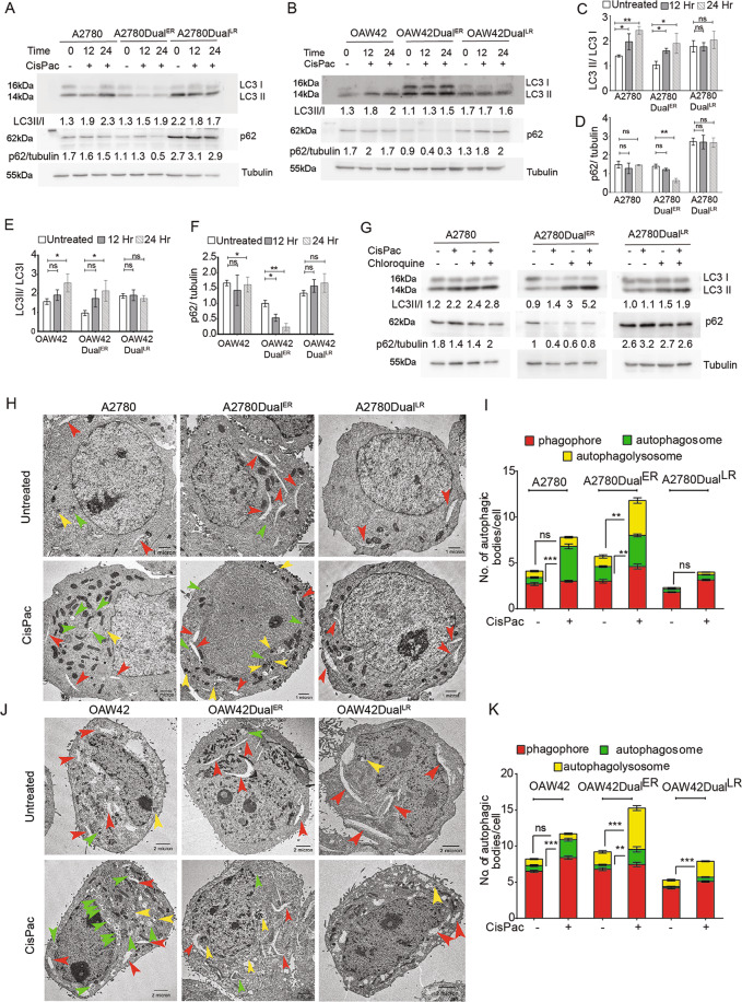
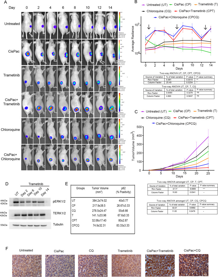
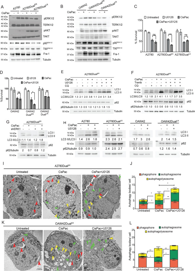
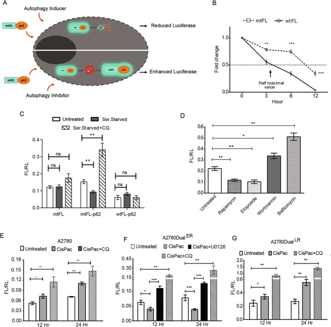
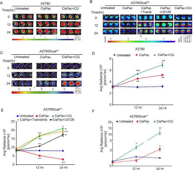
Alterations in key kinases and signaling pathways can fine-tune autophagic flux to promote the development of chemoresistance. Despite empirical evidences of strong association between enhanced autophagic flux with acquired chemoresistance, it is still not understood whether an ongoing autophagic flux is required for both initiation, as well as maintenance of chemoresistance, or is sufficient for one of the either steps. Utilizing indigenously developed cisplatin-paclitaxel-resistant models of ovarian cancer cells, we report an intriguing oscillation in chemotherapy-induced autophagic flux across stages of resistance, which was found to be specifically elevated at the early stages or onset of chemoresistance. Conversely, the sensitive cells and cells at late stages of resistance showed stalled and reduced autophagic flux. This increased flux at early stages of resistance was found to be dictated by a hyperactive ERK1/2 signaling, which when inhibited either pharmacologically (U0126/Trametinib) or genetically, reduced p62 degradation, number of LC3LAMP1 puncta, autophagolysosome formation, and led to chemo-sensitization and apoptosis. Inhibition of ERK1/2 activation also altered the level of UVRAG and Rab7, the two key proteins involved in autophagosome-lysosome fusion. Noninvasive imaging of autophagic flux using a novel autophagy sensor (mtFL-p62 fusion reporter) showed that combinatorial treatment of platinum-taxol along with Trametinib/chloroquine blocked autophagic flux in live cells and tumor xenografts. Interestingly, Trametinib was found to be equally effective in blocking autophagic flux as chloroquine both in live cells and tumor xenografts. Combinatorial treatment of Trametinib and platinum-taxol significantly reduced tumor growth. This is probably the first report of real-time monitoring of chemotherapy-induced autophagy kinetics through noninvasive bioluminescence imaging in preclinical mouse model. Altogether our data suggest that an activated ERK1/2 supports proper completion of autophagic flux at the onset of chemoresistance to endure initial chemotherapeutic insult and foster the development of a highly chemoresistant phenotype, where autophagy becomes dispensable.
关键激酶和信号通路的改变可以微调自噬通量,促进化疗耐药的发展。尽管有经验证据表明增强的自噬通量与获得性化疗耐药之间存在强烈关联,但仍不清楚持续的自噬通量是否是耐药起始和维持所必需的,或者是否足以完成其中一个步骤。利用自主开发的顺铂-紫杉醇耐药卵巢癌细胞模型,我们报告了一个有趣的现象,即在耐药过程中,化疗诱导的自噬通量会发生波动,这种波动在耐药的早期或起始阶段会明显升高。相反,敏感细胞和耐药晚期的细胞显示出停滞和减少的自噬通量。研究发现,耐药早期的这种通量增加是由 ERK1/2 信号的过度活跃所决定的,通过药理学(U0126/Trametinib)或遗传抑制该信号,可减少 p62 的降解、LC3LAMP1 斑点的数量、自噬溶酶体的形成,并导致化疗敏感性和细胞凋亡。抑制 ERK1/2 的激活也改变了 UVRAG 和 Rab7 的水平,这两种关键蛋白参与自噬体-溶酶体融合。使用新型自噬传感器(mtFL-p62 融合报告基因)对自噬通量进行非侵入性成像显示,顺铂-紫杉醇联合 Trametinib/氯喹联合治疗可阻断活细胞和肿瘤异种移植物中的自噬通量。有趣的是,在活细胞和肿瘤异种移植物中,Trametinib 阻断自噬通量的效果与氯喹相当。Trametinib 与顺铂-紫杉醇联合治疗显著抑制肿瘤生长。这可能是首次通过临床前小鼠模型中的非侵入性生物发光成像实时监测化疗诱导的自噬动力学的报道。总之,我们的数据表明,在化疗耐药的起始阶段,激活的 ERK1/2 支持适当完成自噬通量,以耐受初始化疗损伤,并促进高度耐药表型的发展,此时自噬变得可有可无。