Institute of Diagnostic and Interventional Radiology, University Hospital Zurich, Faculty of Medicine, University of Zurich, Raemistrasse 100, 8091, Zurich, Switzerland.
Department of Trauma, University Hospital Zurich, Faculty of Medicine, University of Zurich, 8091, Zurich, Switzerland.
BMC Med Imaging. 2021 Feb 15;21(1):29. doi: 10.1186/s12880-021-00554-y.
CT artifacts induced by orthopedic implants can limit image quality and diagnostic yield. As a number of different strategies to reduce artifact extent exist, the aim of this study was to systematically compare ex vivo the impact of different CT metal artifact reduction (MAR) strategies on spine implants made of either standard titanium or carbon-fiber-reinforced-polyetheretherketone (CFR-PEEK).
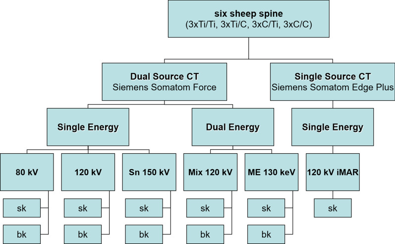
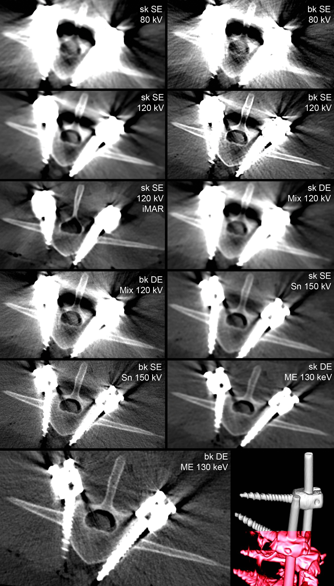
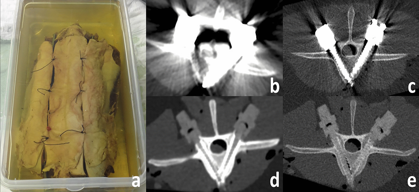
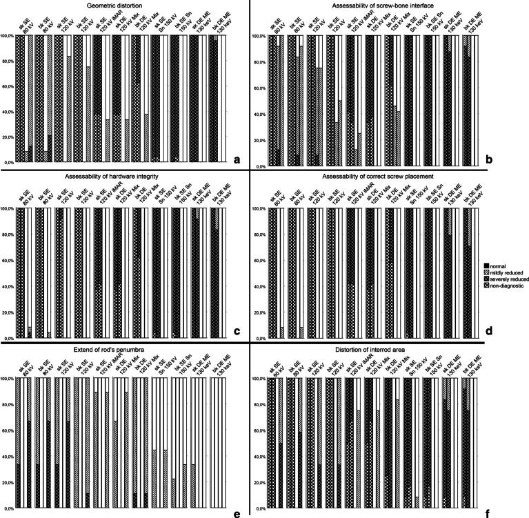
Spine surgeons fluoroscopically-guided prepared six sheep spine cadavers with pedicle screws and rods of either titanium or CFR-PEEK. Samples were subjected to single- and dual-energy (DE) CT-imaging. Different tube voltages (80, DE mixed, 120 and tin-filtered 150 kVp) at comparable radiation dose and iterative reconstruction versus monoenergetic extrapolation (ME) techniques were compared. Also, the influence of image reconstruction kernels (soft vs. bone tissue) was investigated. Qualitative (Likert scores) and quantitative parameters (attenuation changes induced by implant artifact, implant diameter and image noise) were evaluated by two independent radiologists. Artifact degree of different MAR-strategies and implant materials were compared by multiple ANOVA analysis.
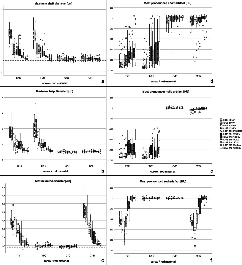
CFR-PEEK implants induced markedly less artifacts than standard titanium implants (p < .001). This effect was substantially larger than any other tested MAR technique. Reconstruction algorithms had small impact in CFR-PEEK implants and differed significantly in MAR efficiency (p < .001) with best MAR performance for DECT ME 130 keV (bone kernel). Significant differences in image noise between reconstruction kernels were seen (p < .001) with minor impact on artifact degree.
CFR-PEEK spine implants induce significantly less artifacts than standard titanium compositions with higher MAR efficiency than any alternate scanning or image reconstruction strategy. DECT ME 130 keV image reconstructions showed least metal artifacts. Reconstruction kernels primarily modulate image noise with minor impact on artifact degree.
骨科植入物引起的 CT 伪影会限制图像质量和诊断效果。由于存在许多不同的降低伪影程度的策略,本研究的目的是系统地比较不同 CT 金属伪影降低(MAR)策略对标准钛和碳纤维增强聚醚醚酮(CFR-PEEK)制成的脊柱植入物的体外影响。
脊柱外科医生在六具羊脊柱尸体标本上经皮椎弓根螺钉和棒进行透视引导准备,植入物分别为钛或 CFR-PEEK 材料。标本进行单次和双能(DE)CT 成像。在相同辐射剂量和迭代重建与单能外推(ME)技术下,比较不同管电压(80、DE 混合、120 和锡过滤 150 kVp)。还研究了图像重建核(软组织与骨组织)的影响。两位独立的放射科医生评估了定性(Likert 评分)和定量参数(植入物伪影引起的衰减变化、植入物直径和图像噪声)。通过多因素方差分析比较了不同 MAR 策略和植入物材料的伪影程度。
CFR-PEEK 植入物引起的伪影明显少于标准钛植入物(p<0.001)。这种效果明显大于任何其他测试的 MAR 技术。重建算法在 CFR-PEEK 植入物中的影响较小,在 MAR 效率上差异显著(p<0.001),DECT ME 130keV(骨核)的 MAR 性能最佳。重建核之间的图像噪声存在显著差异(p<0.001),但对伪影程度的影响较小。
与标准钛成分相比,CFR-PEEK 脊柱植入物引起的伪影明显减少,且比任何其他替代扫描或图像重建策略的 MAR 效率更高。DECT ME 130keV 图像重建显示出最少的金属伪影。重建核主要调节图像噪声,对伪影程度的影响较小。