Pharmaceutical and Biomedical Sciences, 450 College of Pharmacy South, University of Georgia, Athens, GA, 30602, USA.
Proteomics and Mass Spectrometry Facility (PAMS), Department of Chemistry, University of Georgia, Athens, GA, USA.
Lipids Health Dis. 2021 Feb 17;20(1):15. doi: 10.1186/s12944-021-01437-5.
The association of circulating lipids with clinical outcomes of drug-resistant castration-resistant prostate cancer (DR-CRPC) is not fully understood. While it is known that increases in select lipids correlate to decreased survival, neither the mechanisms mediating these alterations nor the correlation of resistance to drug treatments is well characterized.
This gap-in-knowledge was addressed using in vitro models of non-cancerous, hormone-sensitive, CRPC and drug-resistant cell lines combined with quantitative LC-ESI-Orbitrap-MS (LC-ESI-MS/MS) lipidomic analysis and subsequent analysis such as Metaboanalyst and Lipid Pathway Enrichment Analysis (LIPEA).
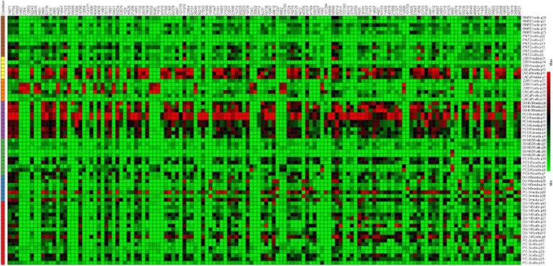
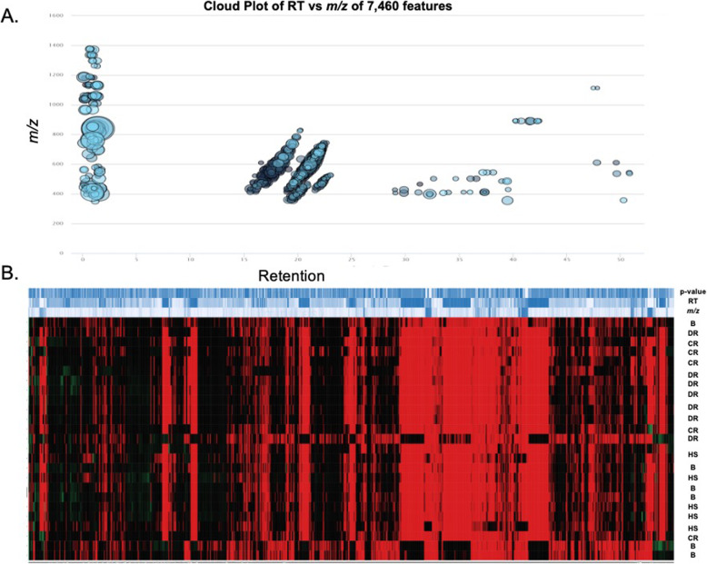
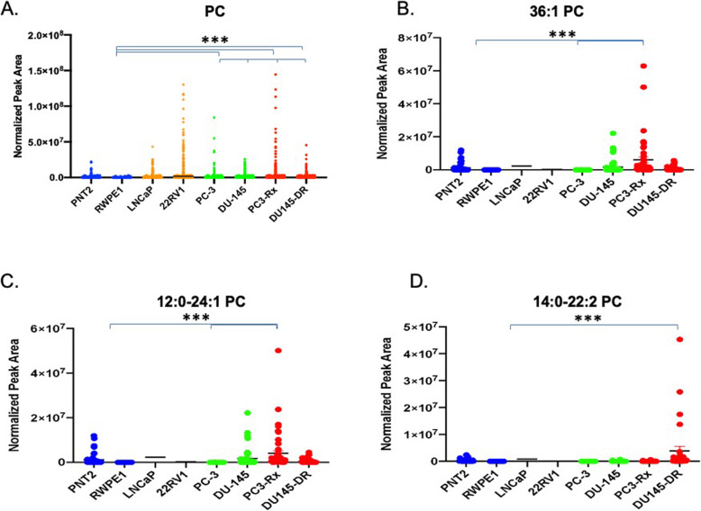
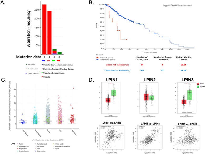
Several lipid regulatory pathways were identified that are associated with Docetaxel resistance in prostate cancer (PCa). These included those controlling glycerophospholipid metabolism, sphingolipid signaling and ferroptosis. In total, 7460 features were identified as being dysregulated between the cell lines studied, and 21 lipid species were significantly altered in drug-resistant cell lines as compared to nonresistant cell lines. Docetaxel resistance cells (PC3-Rx and DU145-DR) had higher levels of phosphatidylcholine (PC), oxidized lipid species, phosphatidylethanolamine (PE), and sphingomyelin (SM) as compared to parent control cells (PC-3 and DU-145). Alterations were also identified in the levels of phosphatidic acid (PA) and diacylglyceride (DAG), whose levels are regulated by Lipin (LPIN), a phosphatidic acid phosphatase that converts PA to DAG. Data derived from cBioPortal demonstrated a population of PCa patients expressing mutations aligning with amplification of LPIN1, LPIN2 and LPIN3 genes. Lipin amplification in these genes correlated to decreased survival in these patients. Lipin-1 mRNA expression also showed a similar trend in PCa patient data. Lipin-1, but not Lipin-2 or - 3, was detected in several prostate cancer cells, and was increased in 22RV1 and PC-3 cell lines. The increased expression of Lipin-1 in these cells correlated with the level of PA.
These data identify lipids whose levels may correlate to Docetaxel sensitivity and progression of PCa. The data also suggest a correlation between the expression of Lipin-1 in cells and patients with regards to prostate cancer cell aggressiveness and patient survivability. Ultimately, these data may be useful for identifying markers of lethal and/or metastatic prostate cancer.
目前尚不完全清楚循环脂质与耐药性去势抵抗性前列腺癌(DR-CRPC)的临床结局之间的关联。虽然已知某些脂质的增加与存活率降低有关,但介导这些变化的机制以及对药物治疗的耐药性的相关性都没有很好地描述。
为了解决这一知识空白,我们使用了非癌性、激素敏感的 CRPC 和耐药细胞系的体外模型,结合定量 LC-ESI-Orbitrap-MS(LC-ESI-MS/MS)脂质组学分析以及随后的分析,如 Metaboanalyst 和脂质通路富集分析(LIPEA)。
确定了几个与前列腺癌(PCa)中多西他赛耐药相关的脂质调节途径。这些途径包括控制甘油磷脂代谢、鞘脂信号和铁死亡的途径。总的来说,在所研究的细胞系之间鉴定出 7460 个特征失调,并且与非耐药细胞系相比,21 种脂质在耐药细胞系中发生显著改变。与亲本对照细胞(PC-3 和 DU-145)相比,多西他赛耐药细胞(PC3-Rx 和 DU145-DR)的磷脂酰胆碱(PC)、氧化脂质、磷脂酰乙醇胺(PE)和鞘磷脂(SM)水平更高。还鉴定出磷酸脂酰酸(PA)和二酰基甘油(DAG)水平的改变,其水平受磷酸脂酰酸磷酸酶 Lipin(LPIN)调节,该酶将 PA 转化为 DAG。来自 cBioPortal 的数据表明,一群表达与 LPIN1、LPIN2 和 LPIN3 基因扩增相吻合的 PCa 患者的突变。这些基因的 Lipin 扩增与这些患者的生存率降低相关。在 PCa 患者数据中,Lipin-1mRNA 表达也呈现出类似的趋势。Lipin-1 而不是 Lipin-2 或 Lipin-3 在几种前列腺癌细胞中被检测到,并且在 22RV1 和 PC-3 细胞系中增加。这些细胞中 Lipin-1 的表达增加与 PA 的水平相关。
这些数据确定了一些脂质,其水平可能与多西他赛敏感性和 PCa 的进展相关。这些数据还表明,在细胞和患者中,Lipin-1 的表达与前列腺癌细胞的侵袭性和患者的存活率之间存在相关性。最终,这些数据可能有助于识别致命性和/或转移性前列腺癌的标志物。