Nuffield Department of Surgical Sciences, John Radcliffe Hospital, University of Oxford, Oxford, OX3 9DU, UK.
Institute of Biomedical Engineering, University of Oxford, Old Road Campus Research Building, Oxford, OX3 7DQ, UK.
Sci Rep. 2021 Feb 23;11(1):4404. doi: 10.1038/s41598-021-83845-2.
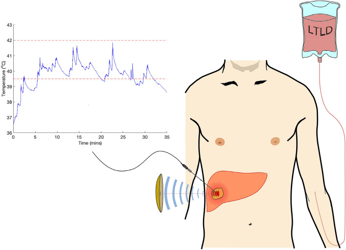
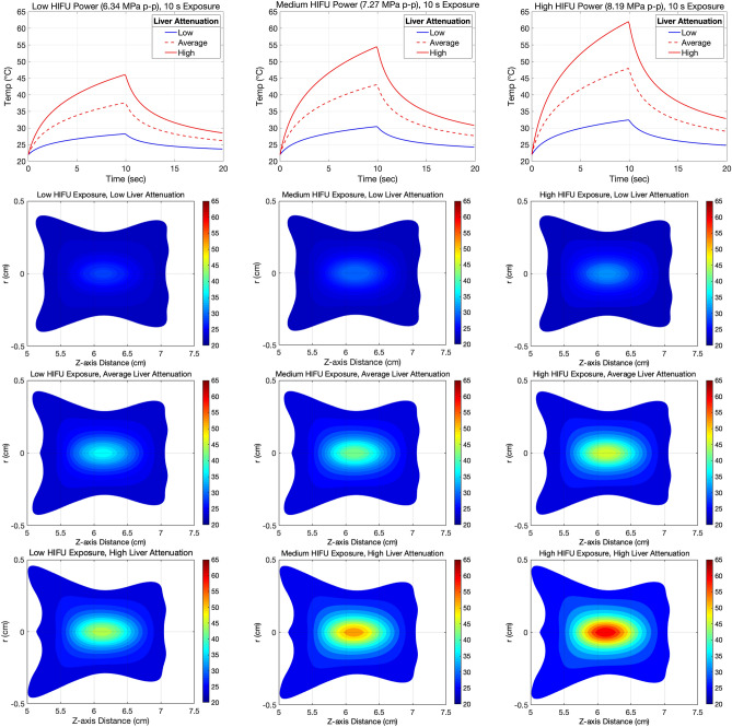
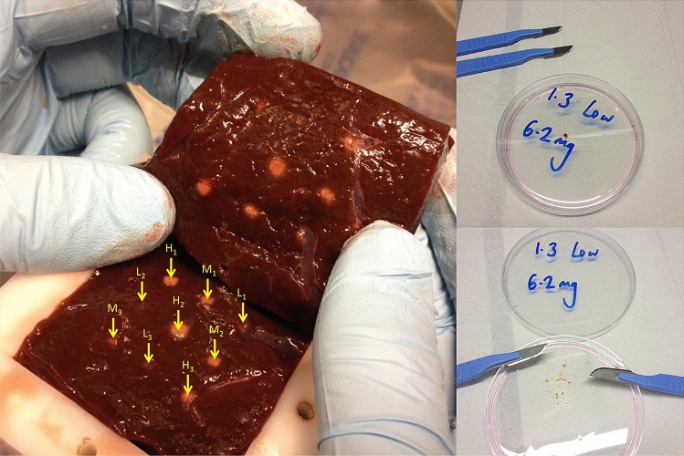
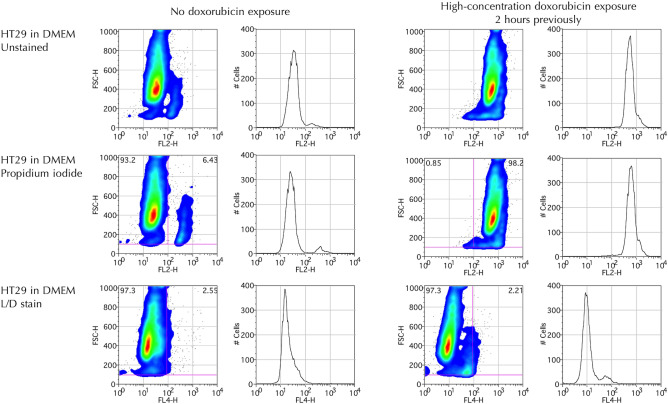
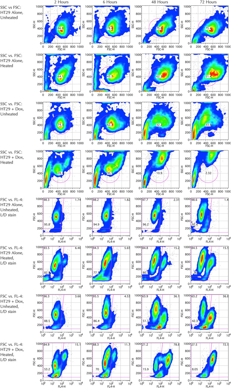
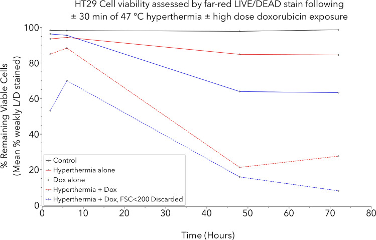
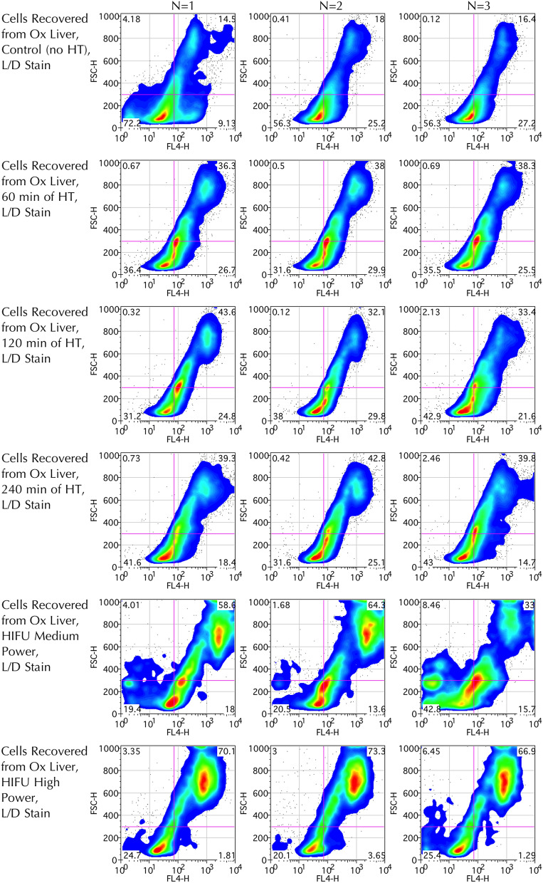
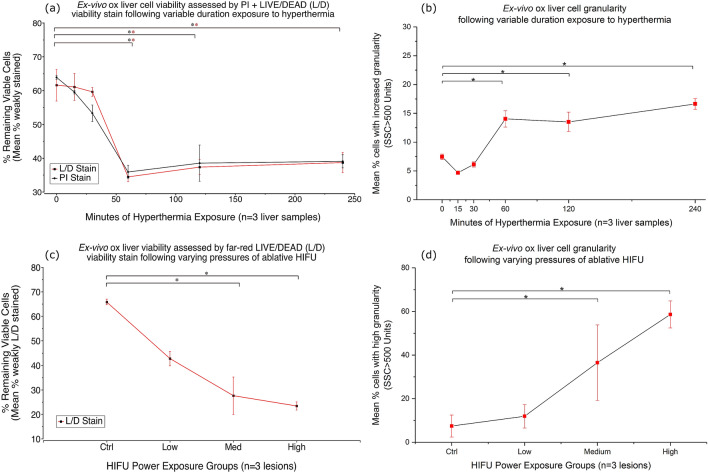
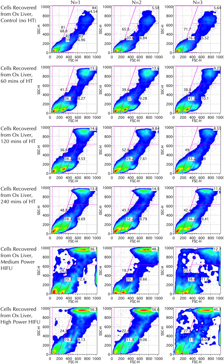
Triggered release and targeted drug delivery of potent anti-cancer agents using hyperthermia-mediated focused-ultrasound (FUS) is gaining momentum in the clinical setting. In early phase studies, tissue biopsy samples may be harvested to assess drug delivery efficacy and demonstrate lack of instantaneous cell death due to FUS exposure. We present an optimised tissue cell recovery method and a cell viability assay, compatible with intra-cellular doxorubicin. Flow cytometry was used to determine levels of cell death with suspensions comprised of: (i) HT29 cell line exposed to hyperthermia (30 min at 47 °C) and/or doxorubicin, or ex-vivo bovine liver tissue exposed to (ii) hyperthermia (up to 2 h at 45 °C), or (iii) ablative high intensity FUS (HIFU). Flow cytometric analysis revealed maximal cell death in HT29 receiving both heat and doxorubicin insults and increases in both cell granularity (p < 0.01) and cell death (p < 0.01) in cells recovered from ex-vivo liver tissue exposed to hyperthermia and high pressures of HIFU (8.2 MPa peak-to-peak free-field at 1 MHz) relative to controls. Ex-vivo results were validated with microscopy using pan-cytokeratin stain. This rapid, sensitive and highly quantitative cell-viability method is applicable to the small masses of liver tissue typically recovered from a standard core biopsy (5-20 mg) and may be applied to tissues of other histological origins including immunostaining.
使用热疗介导的聚焦超声(FUS)实现强效抗癌药物的触发释放和靶向药物输送,在临床环境中正逐渐受到关注。在早期研究阶段,可以采集组织活检样本,以评估药物输送效果,并证明由于 FUS 暴露而没有即时的细胞死亡。我们提出了一种优化的组织细胞回收方法和细胞活力测定法,该方法与细胞内阿霉素兼容。采用流式细胞术来确定细胞死亡水平,悬浮液包含:(i)暴露于热疗(47°C 下 30 分钟)和/或阿霉素的 HT29 细胞系,或(ii)暴露于热疗(最高 45°C 下 2 小时)的牛肝组织,或(iii)消融高强度 FUS(HIFU)。流式细胞术分析显示,同时接受热疗和阿霉素刺激的 HT29 细胞死亡最多,且从暴露于热疗和 HIFU 高压(1MHz 时峰值-峰值自由场 8.2MPa)的离体肝组织中回收的细胞的细胞颗粒度(p<0.01)和细胞死亡(p<0.01)均增加。离体实验结果通过使用广谱细胞角蛋白染色的显微镜检查得到验证。这种快速、灵敏且高度定量的细胞活力测定方法适用于从标准核心活检中通常回收的小肝组织质量(5-20mg),并且可应用于包括免疫染色在内的其他组织起源的组织。