Biotechnology Research and Training Center, University of North Carolina-Pembroke, Pembroke, NC, USA.
U.S. Army Research Laboratory, Aberdeen Proving Ground, MD, USA.
Brain Pathol. 2021 May;31(3):e12936. doi: 10.1111/bpa.12936. Epub 2021 Feb 24.
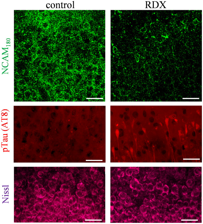
Explosive shockwaves, and other types of blast exposures, are linked to injuries commonly associated with military service and to an increased risk for the onset of dementia. Neurological complications following a blast injury, including depression, anxiety, and memory problems, often persist even when brain damage is undetectable. Here, hippocampal explants were exposed to the explosive 1,3,5-trinitro-1,3,5-triazinane (RDX) to identify indicators of blast-induced changes within important neuronal circuitries. Highly controlled detonations of small, 1.7-gram RDX spherical charges reduced synaptic markers known to be downregulated in cognitive disorders, but without causing overt neuronal loss or astroglial responses. In the absence of neuromorphological alterations, levels of synaptophysin, GluA1, and synapsin IIb were significantly diminished within 24 hr, and these synaptic components exhibited progressive reductions following blast exposure as compared to their stable maintenance in control explants. In contrast, labeling of the synapsin IIa isoform remained unaltered, while neuropilar staining of other markers decreased, including synapsin IIb and neural cell adhesion molecule (NCAM) isoforms, along with evidence of NCAM proteolytic breakdown. NCAM displayed a distinct decline after the RDX blasts, whereas NCAM and NCAM exhibited smaller or no deterioration, respectively. Interestingly, the extent of synaptic marker reduction correlated with AT8-positive tau levels, with tau pathology stochastically found in CA1 neurons and their dendrites. The decline in synaptic components was also reflected in the size of evoked postsynaptic currents recorded from CA1 pyramidals, which exhibited a severe and selective reduction. The identified indicators of blast-mediated synaptopathy point to the need for early biomarkers of explosives altering synaptic integrity with links to dementia risk, to advance strategies for both cognitive health and therapeutic monitoring.
爆炸冲击波和其他类型的爆炸暴露与常见的军事服务相关的损伤以及痴呆症发病风险增加有关。爆炸伤后的神经并发症,包括抑郁、焦虑和记忆问题,即使在脑损伤无法检测到时也常常持续存在。在这里,海马体外植体暴露于爆炸物 1,3,5-三硝基-1,3,5-三嗪烷(RDX)下,以确定重要神经元回路中爆炸诱导变化的指标。通过对小的 1.7 克 RDX 球形炸药进行高度受控的爆炸,可以减少已知在认知障碍中下调的突触标志物,但不会导致明显的神经元丢失或星形胶质细胞反应。在没有神经形态学改变的情况下,突触小体蛋白、GluA1 和突触素 IIb 的水平在 24 小时内显著降低,并且这些突触成分在爆炸暴露后表现出逐渐减少,而在对照外植体中则稳定维持。相比之下,突触素 IIa 同工型的标记保持不变,而其他标记物的神经突染色减少,包括突触素 IIb 和神经细胞黏附分子(NCAM)同工型,以及 NCAM 蛋白水解断裂的证据。NCAM 在 RDX 爆炸后表现出明显的下降,而 NCAM 和 NCAM 的下降较小或没有下降。有趣的是,突触标记物减少的程度与 AT8 阳性 tau 水平相关,tau 病理学随机存在于 CA1 神经元及其树突中。突触成分的减少也反映在从 CA1 锥体记录的诱发突触后电流的大小上,其表现出严重和选择性的减少。鉴定出的爆炸介导的突触病变指标表明,需要早期的爆炸物改变突触完整性的生物标志物与痴呆风险相关联,以推进认知健康和治疗监测的策略。