Department of General Ophthalmology, Medical University of Lublin, Lublin, Poland.
Departament of Mathematics and Medical Biostatistics, Medical University of Lublin, Lublin, Poland.
PLoS One. 2021 Feb 25;16(2):e0247399. doi: 10.1371/journal.pone.0247399. eCollection 2021.
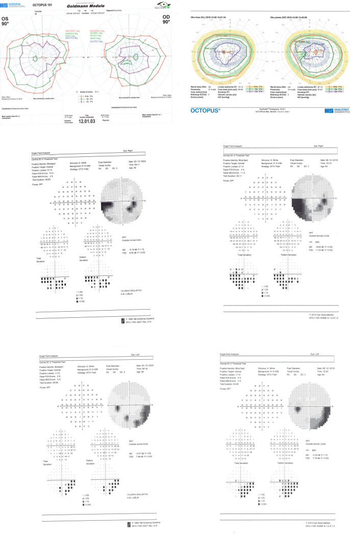
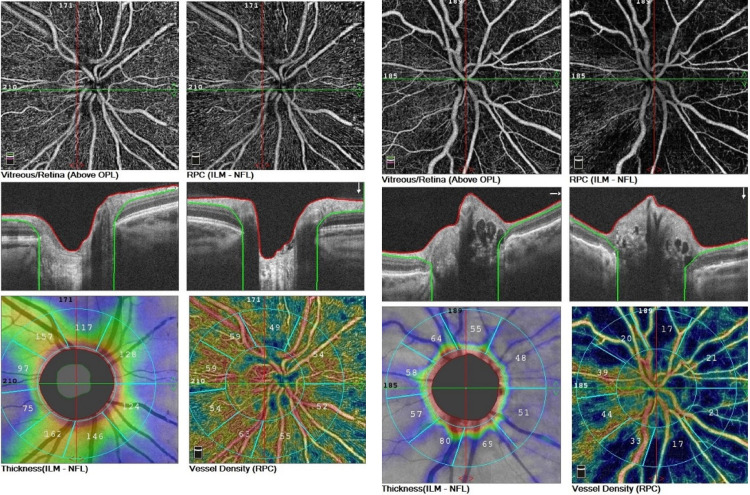
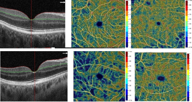
The aim of the study is to evaluate the progression of visual field (VF) defects over 16 years of observation and to assess abnormalities in vessels and retinal nerve fibre layer (RNFL) thickness in patients with optic disc drusen (ODD). Both static automated perimetry (SAP) and semi-automated kinetic perimetry (SKP) were performed in 16 eyes of 8 patients (mean age 54 years) with ODD among 26 eyes of 13 patients examined 16 years before. The area of I2e, I4e, III4e, and V4e isopters was measured in deg2. The MD and PSD parameters were estimated using SAP. Optical coherence tomography angiography (OCT-A) was additionally performed in 16 ODD eyes and 16 eyes of 8 healthy subjects to estimate the RNFL thickness and vessel density of the optic nerve disc and the macula. The differences in all isopter areas of SKP and SAP parameters after 16 years were not significant. The analysis of OCT-A showed a significant reduction of the vessel density and RNFL of the peripapillary area in each segment in patients with ODD, compared with the control group. The highest reduction of RNFL was observed in the superior segment of the optic disc area (92.56μm vs 126.63μm) also the macular thickness was decreased in ODD patients, compared with the control group. In the macula, there was a significant vascular defect in the whole superficial layer and in the parafoveal deep layer. A strong significant correlation of the parafoveal deep plexus with MD and PSD parameters was detected. In conclusion, VF loss due to ODD after 16 years of the follow-up was not significant both in SKP and SAP. ODD caused a reduced vessel density and RNFL, as well as macular thickness in OCT-A. SAP parameters were influenced by parafoveal deep plexus.
本研究旨在评估视盘玻璃膜疣(ODD)患者在 16 年的观察期间视野(VF)缺损的进展,并评估血管和视网膜神经纤维层(RNFL)厚度的异常。在 26 只眼中,对 13 例患者的 16 年前检查的 26 只眼中的 8 例患者(平均年龄 54 岁)的 16 只眼进行了静态自动视野计(SAP)和半自动动态视野计(SKP)检查。在 deg2 中测量 I2e、I4e、III4e 和 V4e 等视线的面积。使用 SAP 估计 MD 和 PSD 参数。在 16 只 ODD 眼中和 8 名健康受试者的 16 只眼中进一步进行光学相干断层扫描血管造影(OCT-A),以估计视盘和黄斑的 RNFL 厚度和血管密度。16 年后,SKP 和 SAP 参数的所有等视线区域的差异均不显著。OCT-A 分析显示,与对照组相比,ODD 患者每个节段的视盘和黄斑周围区域的血管密度和 RNFL 显著降低。与对照组相比,ODD 患者的视盘区域的上节段的 RNFL 降低最大(92.56μm 对 126.63μm),黄斑厚度也降低。在黄斑区,整个浅层和旁中心深层均存在明显的血管缺损。在旁中心深层丛中检测到与 MD 和 PSD 参数有很强的显著相关性。结论:在 16 年的随访中,SKP 和 SAP 中由于 ODD 导致的 VF 损失并不显著。ODD 在 OCT-A 中引起血管密度和 RNFL 以及黄斑厚度降低。SAP 参数受旁中心深层丛的影响。