Wang Songjian, Lin Meng, Sun Liwei, Chen Xueqing, Fu Xinxing, Yan LiLi, Li Chunlin, Zhang Xu
School of Biomedical Engineering, Capital Medical University, Beijing, China.
Beijing Key Laboratory of Fundamental Research on Biomechanics in Clinical Application, Capital Medical University, Beijing, China.
Front Neurosci. 2021 Feb 5;14:624484. doi: 10.3389/fnins.2020.624484. eCollection 2020.
Patients with severe profound hearing loss could benefit from cochlear implantation (CI). However, the neural mechanism of such benefit is still unclear. Therefore, we analyzed the electroencephalogram (EEG) and behavioral indicators of auditory function remodeling in patients with CI. Both indicators were sampled at multiple time points after implantation (1, 90, and 180 days).
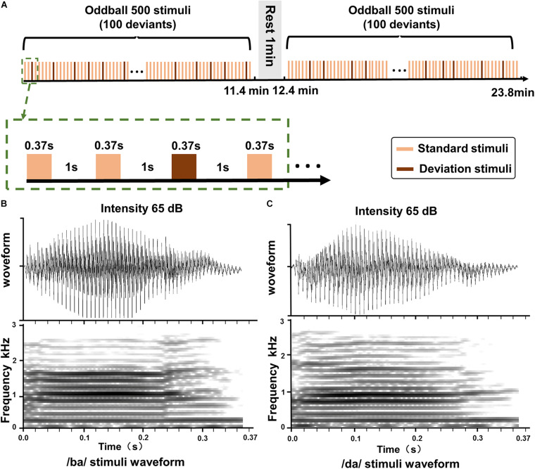
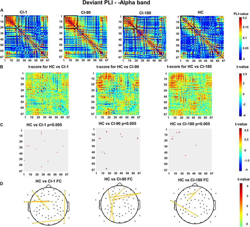
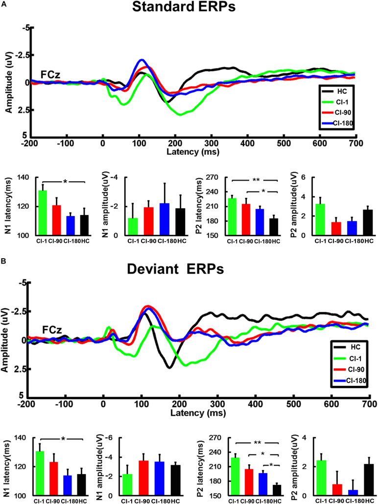
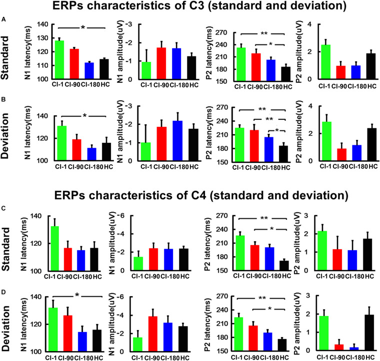
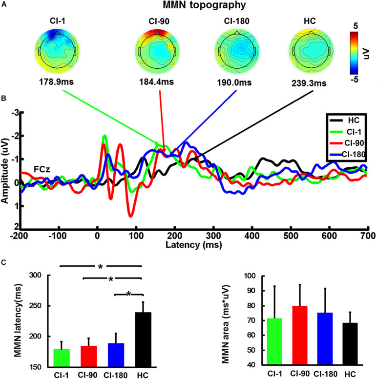
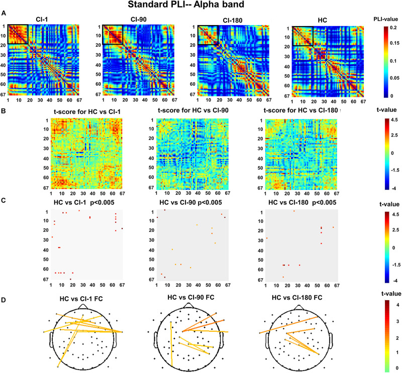
First, the speech perception ability was evaluated with the recording of a list of Chinese words and sentences in 15 healthy controls (HC group) and 10 patients with CI (CI group). EEG data were collected using an oddball paradigm. Then, the characteristics of event-related potentials (ERPs) and mismatch negative (MMN) were compared between the CI group and the HC group. In addition, we analyzed the phase lag indices (PLI) in the CI group and the HC group and calculated the difference in functional connectivity between the two groups at different stages after implantation.
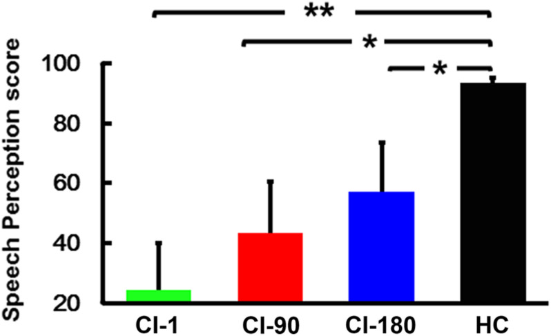
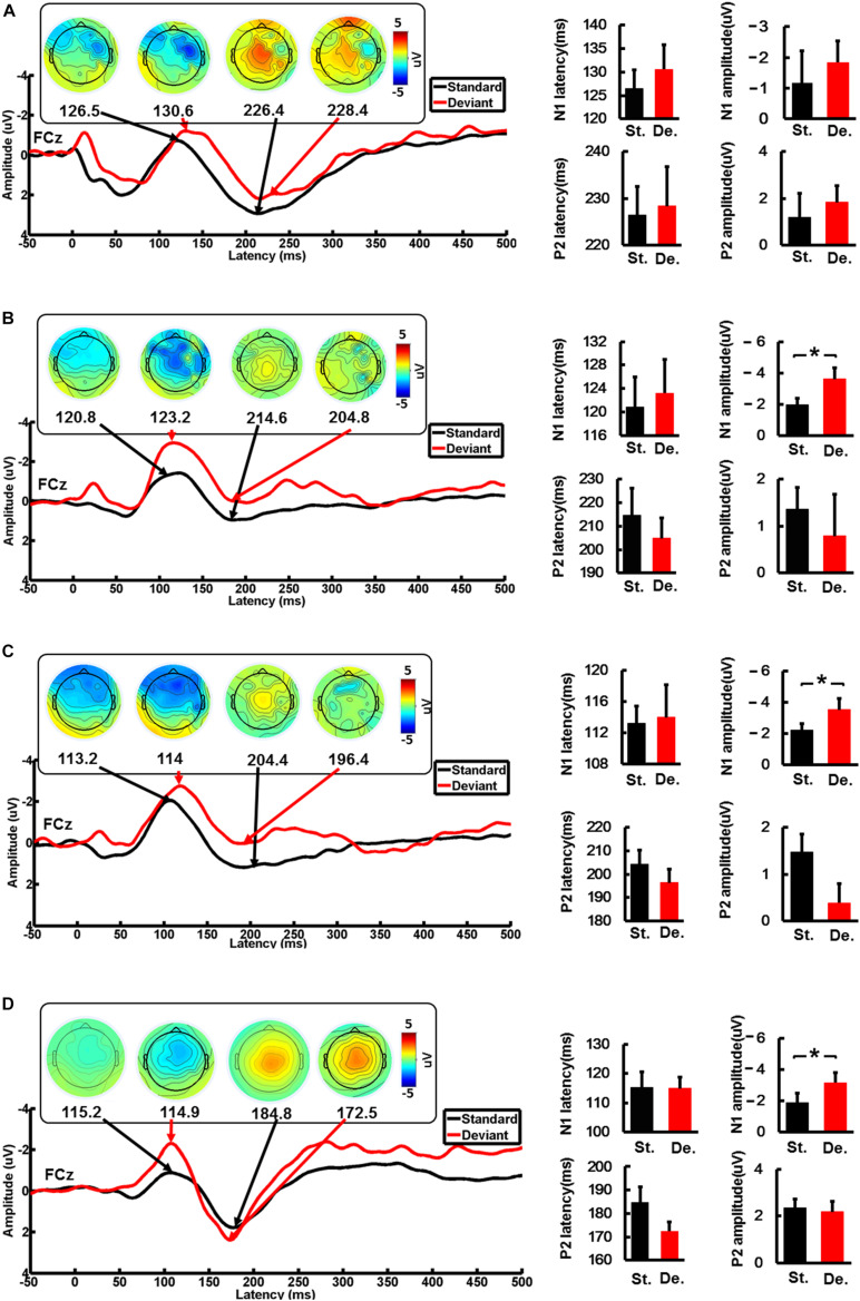
The behavioral indicator, speech recognition ability, in CI patients improved as the implantation time increased. The MMN analysis showed that CI patients could recognize the difference between standard and deviation stimuli just like the HCs 90 days after cochlear implantation. Comparing the latencies of N1/P2/MMN between the CI group and the HC group, we found that the latency of N1/P2 in CI patients was longer, while the latency of MMN in CI users was shorter. In addition, PLI-based whole-brain functional connectivity (PLI-FC) showed that the difference between the CI group and the HC group mainly exists in electrode pairs between the bilateral auditory area and the frontal area. Furthermore, all those differences gradually decreased with the increase in implantation time.
The N1 amplitude, N1/P2/MMN latency, and PLI-FC in the alpha band may reflect the process of auditory function remodeling and could be an objective index for the assessment of speech perception ability and the effect of cochlear implantation.
重度极重度听力损失患者可从人工耳蜗植入(CI)中获益。然而,这种获益的神经机制仍不清楚。因此,我们分析了人工耳蜗植入患者听觉功能重塑的脑电图(EEG)和行为指标。这两个指标均在植入后的多个时间点(1天、90天和180天)进行采样。
首先,通过记录15名健康对照者(HC组)和10名人工耳蜗植入患者(CI组)的中文单词和句子列表来评估言语感知能力。使用oddball范式收集EEG数据。然后,比较CI组和HC组之间事件相关电位(ERP)和失配负波(MMN)的特征。此外,我们分析了CI组和HC组的相位滞后指数(PLI),并计算了植入后不同阶段两组之间功能连接性的差异。
CI患者的行为指标,即言语识别能力,随着植入时间的增加而提高。MMN分析表明,人工耳蜗植入90天后,CI患者能够像健康对照者一样识别标准刺激和偏差刺激之间的差异。比较CI组和HC组之间N1/P2/MMN的潜伏期,我们发现CI患者中N1/P2的潜伏期较长,而CI使用者中MMN的潜伏期较短。此外,基于PLI的全脑功能连接性(PLI-FC)表明,CI组和HC组之间的差异主要存在于双侧听觉区域和额叶之间的电极对中。此外,所有这些差异随着植入时间的增加而逐渐减小。
α波段中的N1波幅、N1/P2/MMN潜伏期和PLI-FC可能反映听觉功能重塑的过程,并且可能是评估言语感知能力和人工耳蜗植入效果的客观指标。