Danish Research Centre for Magnetic Resonance, Centre for Functional and Diagnostic Imaging and Research, Copenhagen University Hospital Hvidovre, 2650 Hvidovre, Denmark
Department of Neurosciences, Reproductive Sciences and Odontostomatology, University Federico II of Naples, 80131 Naples, Italy.
J Neurosci. 2021 Apr 7;41(14):3163-3179. doi: 10.1523/JNEUROSCI.0390-20.2021. Epub 2021 Mar 2.
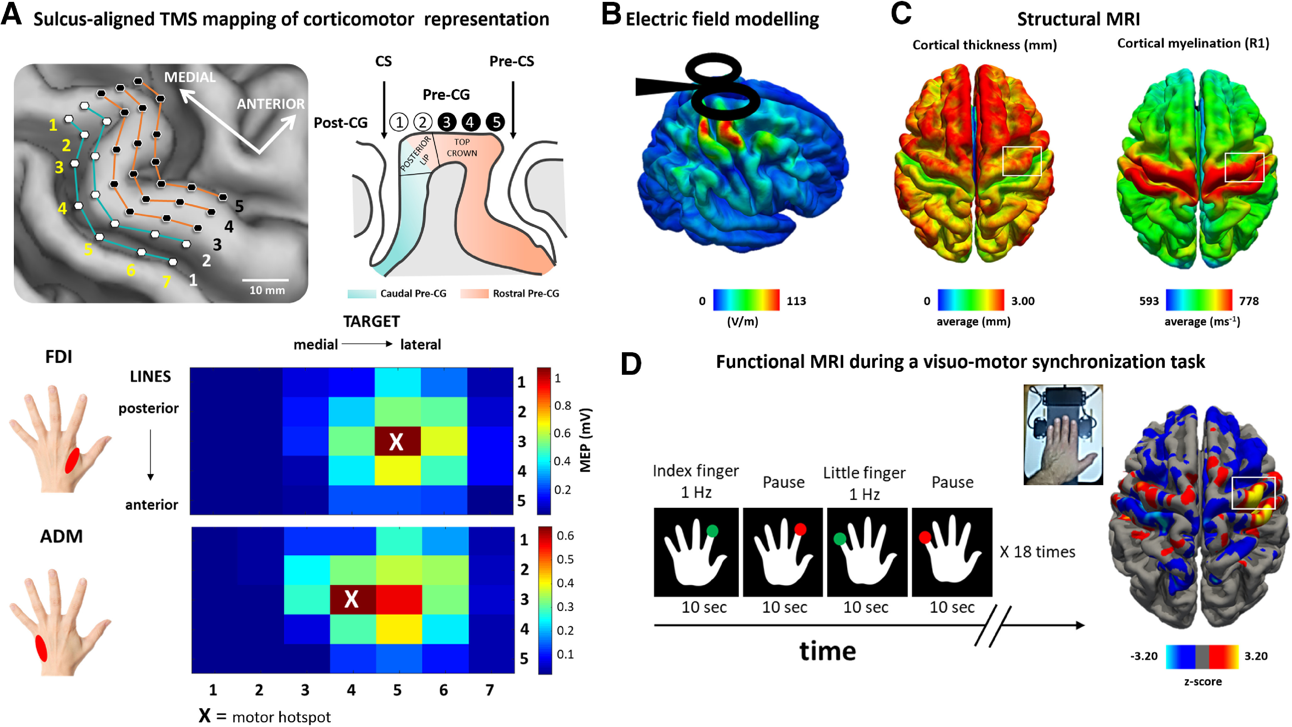
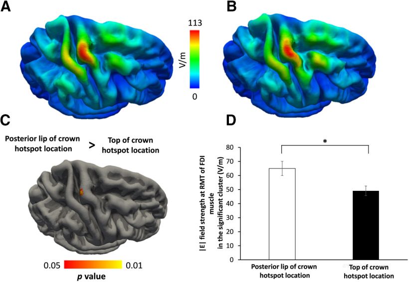
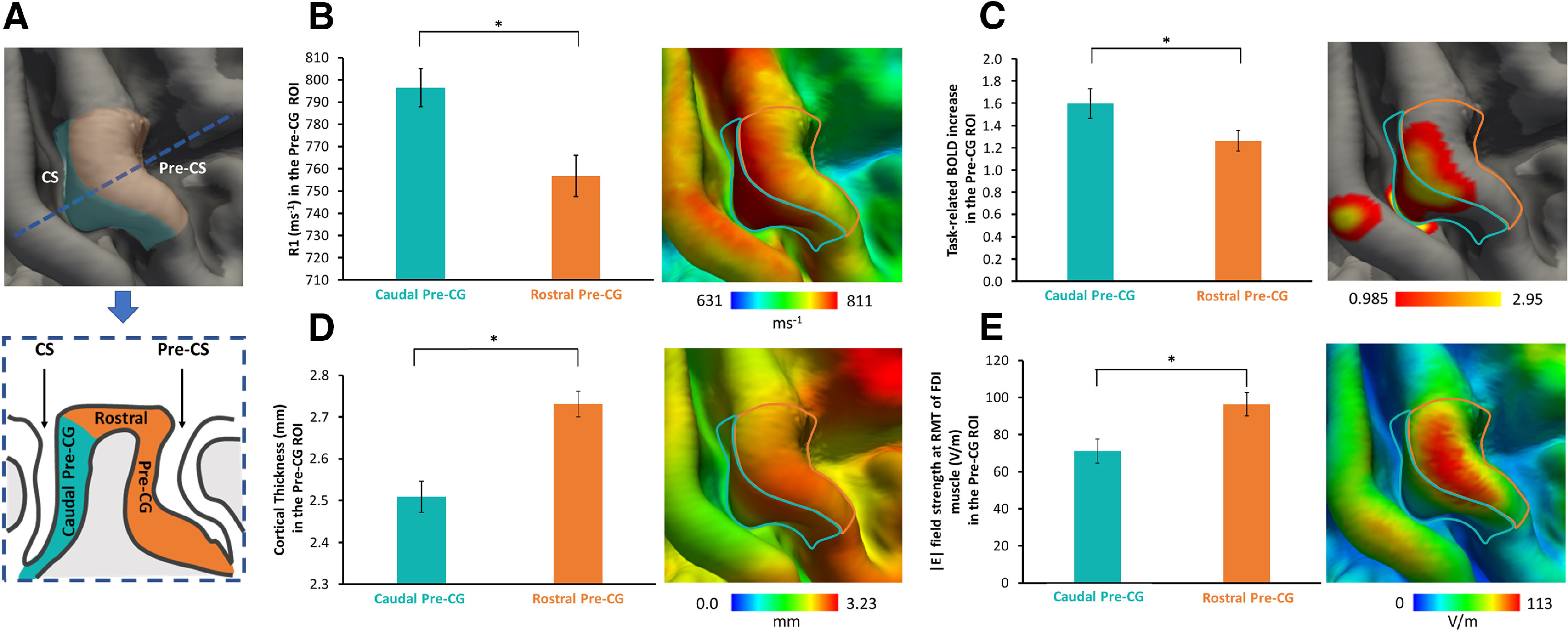
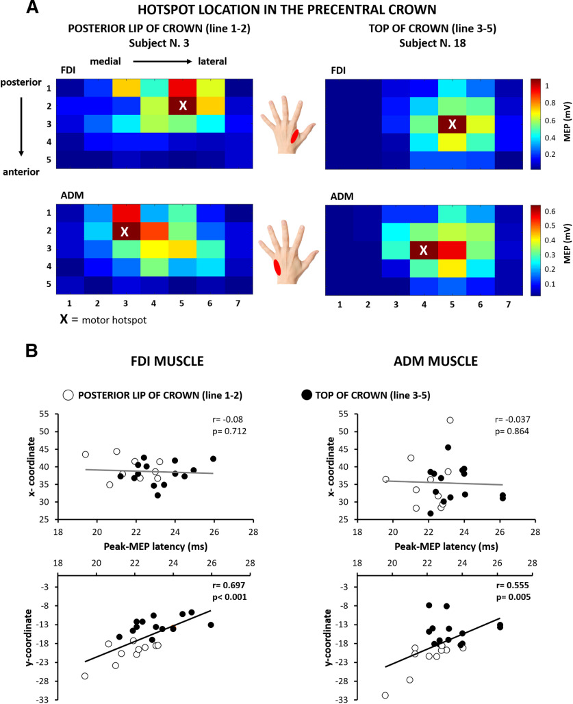
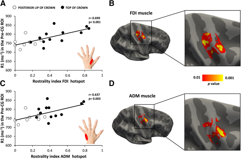
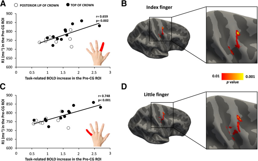
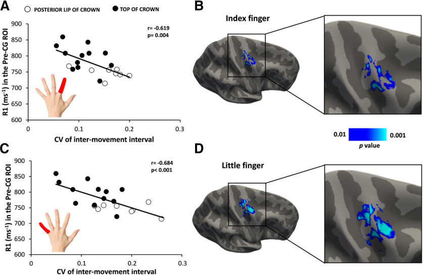
The primary motor cortex hand area (M1) and adjacent dorsal premotor cortex (PMd) form the so-called motor hand knob in the precentral gyrus. M1 and PMd are critical for dexterous hand use and are densely interconnected via corticocortical axons, lacking a sharp demarcating border. In 24 young right-handed volunteers, we performed multimodal mapping to delineate the relationship between structure and function in the right motor hand knob. Quantitative structural magnetic resonance imaging (MRI) at 3 tesla yielded regional R1 maps as a proxy of cortical myelin content. Participants also underwent functional MRI (fMRI). We mapped task-related activation and temporal precision, while they performed a visuomotor synchronization task requiring visually cued abduction movements with the left index or little finger. We also performed sulcus-aligned transcranial magnetic stimulation of the motor hand knob to localize the optimal site (hotspot) for evoking a motor evoked potential (MEP) in two intrinsic hand muscles. Individual motor hotspot locations varied along the rostrocaudal axis. The more rostral the motor hotspot location in the precentral crown, the longer were corticomotor MEP latencies. "Hotspot rostrality" was associated with the regional myelin content in the precentral hand knob. Cortical myelin content also correlated positively with task-related activation of the precentral crown and temporal precision during the visuomotor synchronization task. Together, our results suggest a link among cortical myelination, the spatial cortical representation, and temporal precision of finger movements. We hypothesize that the myelination of cortical axons facilitates neuronal integration in PMd and M1 and, hereby, promotes the precise timing of movements. Here we used magnetic resonance imaging and transcranial magnetic stimulation of the precentral motor hand knob to test for a link among cortical myelin content, functional corticomotor representations, and manual motor control. A higher myelin content of the precentral motor hand knob was associated with more rostral corticomotor presentations, with stronger task-related activation and a higher precision of movement timing during a visuomotor synchronization task. We propose that a high precentral myelin content enables fast and precise neuronal integration in M1 (primary motor cortex) and dorsal premotor cortex, resulting in higher temporal precision during dexterous hand use. Our results identify the degree of myelination as an important structural feature of the neocortex that is tightly linked to the function and behavior supported by the cortical area.
初级运动皮层手部区域(M1)和相邻的背侧运动前皮层(PMd)在中央前回中形成所谓的运动手旋钮。M1 和 PMd 对手部灵巧运动至关重要,并且通过皮质皮质轴突密集地相互连接,没有明显的边界。在 24 名年轻的右利手志愿者中,我们进行了多模态映射,以描绘右侧运动手旋钮的结构与功能之间的关系。在 3 特斯拉的定量结构磁共振成像(MRI)中,获得了区域 R1 图作为皮质髓鞘含量的替代物。参与者还接受了功能磁共振成像(fMRI)。当他们执行视觉运动同步任务(需要用左手食指或小指进行视觉提示外展运动)时,我们绘制了与任务相关的激活和时间精度图。我们还对运动手旋钮进行了沟回对齐的经颅磁刺激,以定位在两个内在手部肌肉中诱发运动诱发电位(MEP)的最佳位置(热点)。个体运动热点位置沿前后轴变化。中央前回冠部的运动热点位置越靠前,皮质运动 MEP 潜伏期越长。“热点前突性”与中央前回手旋钮的区域髓鞘含量相关。皮质髓鞘含量还与视觉运动同步任务期间中央前回冠部的任务相关激活和时间精度呈正相关。总之,我们的结果表明皮质髓鞘、手指运动的空间皮质代表和时间精度之间存在联系。我们假设皮质轴突的髓鞘形成促进了 PMd 和 M1 中的神经元整合,从而促进了运动的精确定时。在这里,我们使用磁共振成像和中央前回运动手旋钮的经颅磁刺激来测试皮质髓鞘含量、功能皮质运动代表和手动运动控制之间的联系。中央前回运动手旋钮的髓鞘含量较高与皮质运动呈现更靠前有关,与视觉运动同步任务期间更强的任务相关激活和更高的运动定时精度有关。我们提出,中央前回高髓鞘含量能够实现 M1(初级运动皮层)和背侧运动前皮层的快速而精确的神经元整合,从而在手灵巧运动中实现更高的时间精度。我们的结果确定了髓鞘程度作为新皮层的重要结构特征,它与皮层区域支持的功能和行为紧密相关。