Patra Aparna, Thakkar Pratibha S, Makhoul Majd, Bada Henrietta S
Division of Neonatology, Department of Pediatrics, Kentucky Children's Hospital, University of Kentucky, Lexington, KY, United States.
Division of Pediatric Cardiology, Department of Pediatrics, Kentucky Children's Hospital, University of Kentucky, Lexington, KY, United States.
Front Pediatr. 2021 Feb 25;9:648584. doi: 10.3389/fped.2021.648584. eCollection 2021.
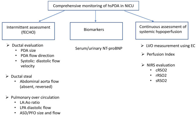
Delay in closure of ductus arteriosus in postnatal life may lead to serious consequences and complications in an extremely premature neonate secondary to hemodynamic alterations in regional blood flow pattern in various organs. Despite the widespread recognition amongst neonatologists to identify a hemodynamically significant patent ductus arteriosus (hsPDA) early in the postnatal course, there is lack of consensus in its definition and thus the threshold to initiate treatment. Echocardiographic assessment of PDA shunt size and volume combined with neonatologists' impression of clinical significance is most frequently used to determine the need for treatment of PDA. Common clinical signs of hsPDA utilized as surrogate for decreased tissue perfusion may lag behind early echocardiographic signs. Although echocardiogram allows direct assessment of PDA shunt and hemodynamic alterations in the heart, it is limited by dependence on pediatric cardiologist availability, interobserver variation and isolated time point assessment. Electrical cardiometry (EC) is a non-invasive continuous real time measurement of cardiac output by applying changes in thoracic electrical impedance. EC has been validated in preterm newborns by concomitant transthoracic echocardiogram assessments and may be beneficial in studying changes in cardiac output in premature newborns with hsPDA. Alterations in perfusion index derived from continuous pulse oximetry monitoring has been used to study changes in cardiac performance and tissue perfusion in infants with PDA. Near infrared spectroscopy (NIRS) has been used to objectively and continuously assess variations in renal, mesenteric, and cerebral oxygen saturation and thus perfusion changes due to diastolic vascular steal from hsPDA in preterm neonates. Doppler ultrasound studies measuring resistive indices in cerebral circulation indicate disturbance in cerebral perfusion secondary to ductal steal. With recent trends of change in practice toward less intervention in care of preterm newborn, treatment strategy needs to be targeted for select preterm population most vulnerable to adverse hemodynamic effects of PDA. Integration of these novel ways of hemodynamic and tissue perfusion assessment in routine clinical care may help mitigate the challenges in defining and targeting treatment of hsPDA thereby improving outcomes in extremely premature neonates.
出生后动脉导管延迟关闭可能会给极早产儿带来严重后果及并发症,这是由于各器官局部血流模式的血流动力学改变所致。尽管新生儿科医生普遍认识到要在出生后早期识别出具有血流动力学意义的动脉导管未闭(hsPDA),但其定义以及启动治疗的阈值仍缺乏共识。对动脉导管未闭分流大小和容积进行超声心动图评估,并结合新生儿科医生对临床意义的判断,是最常用于确定动脉导管未闭治疗需求的方法。用作组织灌注减少替代指标的hsPDA常见临床体征可能滞后于早期超声心动图体征。虽然超声心动图可直接评估动脉导管未闭分流及心脏血流动力学改变,但它受限于依赖儿科心脏病专家、观察者间差异以及孤立的时间点评估。心电描记术(EC)是通过应用胸壁电阻抗变化对心输出量进行非侵入性连续实时测量。通过同步经胸超声心动图评估,EC已在早产儿中得到验证,可能有助于研究患有hsPDA的早产儿的心输出量变化。连续脉搏血氧饱和度监测得出的灌注指数变化已用于研究患有动脉导管未闭婴儿的心脏功能和组织灌注变化。近红外光谱(NIRS)已用于客观连续地评估肾、肠系膜和脑氧饱和度的变化,从而评估早产儿因hsPDA舒张期血管窃血导致的灌注变化。测量脑循环阻力指数的多普勒超声研究表明,导管窃血会导致脑灌注紊乱。随着近期临床实践趋势朝着减少对早产儿护理干预的方向发展,治疗策略需要针对最易受PDA不良血流动力学影响的特定早产儿群体。将这些血流动力学和组织灌注评估的新方法整合到常规临床护理中,可能有助于应对定义和靶向治疗hsPDA方面的挑战,从而改善极早产儿的预后。