Liu Jiao, Li Qingtao, Shi Nannan, Chen Yi, Li Yesheng, Zhang Meng, Huang Yangqing
Hepatobiliary Surgery Department Shanghai Public Health Clinical Center Shanghai China.
Interventional Department Shanghai Public Health Clinical Center Shanghai China.
JGH Open. 2021 Feb 18;5(3):382-389. doi: 10.1002/jgh3.12507. eCollection 2021 Mar.
Studies have found that gadolinium-ethoxybenzyl-diethylenetriamine pentaacetic acid (Gd-EOB-DTPA)-enhanced T1 mapping magnetic resonance imaging (MRI) could assess liver fibrosis, cirrhosis, and function with high effectiveness. The aim of this study is to explore the efficacy of MRI in predicting the safety of hepatectomy.
Forty-nine patients who underwent liver resection were recruited. Gd-EOB-DTPAenhanced MRI examination was performed 1 week before surgery, and the rate of T1 relaxation time reduction (ΔT1%) of liver parenchyma was calculated. Posthepatectomy liver failure (PHLF) was defined by the "50-50 criteria" and International Study Group of Liver Surgery (ISGLS) classification, respectively, and posthepatectomy complications (PHC) were defined by the Clavien-Dindo grading system. The effectiveness of ΔT1% in predicting the occurrence of PHLF and PHC was analyzed.
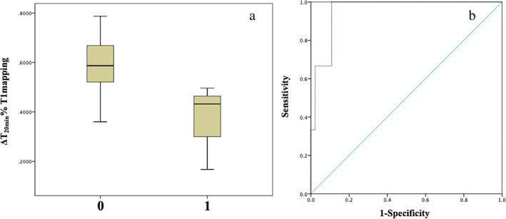
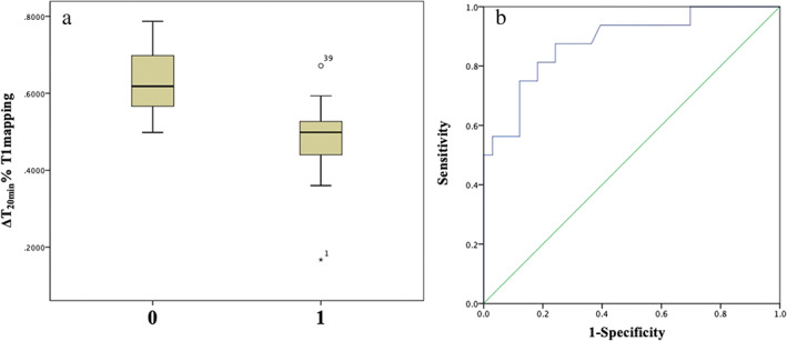
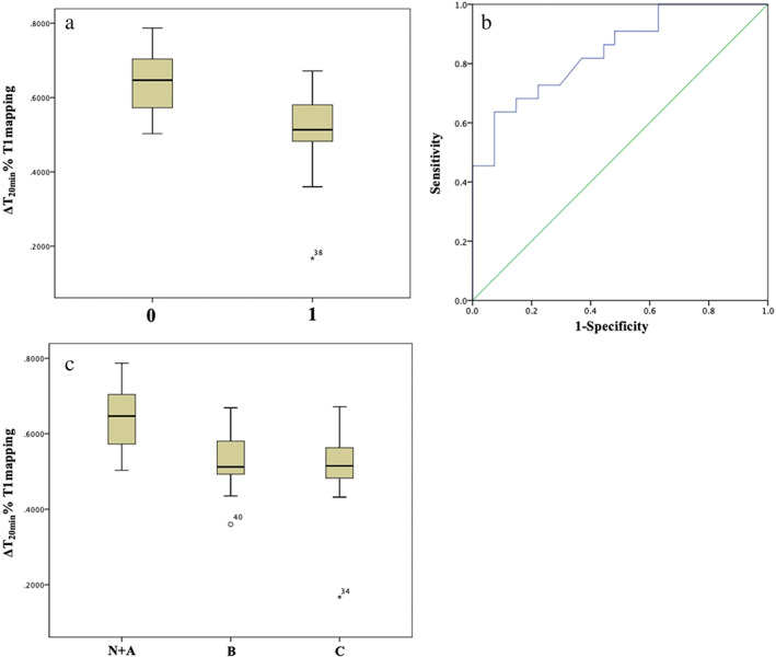
The area under the curve (AUC) for ΔT1% predicting PHLF meeting "50-50 criteria" was 0.957, with a cutoff value of 0.497, sensitivity of 100%, and specificity of 89.1%. The AUC for predicting ISGLS grade B/C (severe) PHLF was 0.84, with a cutoff value of 0.5232, sensitivity of 63.6%, and specificity of 92.6%. The AUC for predicting PHC of Clavien-Dindo grades 3-5 (severe) was 0.882, with a cutoff value of 0.5646, sensitivity of 87.5%, and specificity of 75.8%. Univariate and multivariate analyses showed that ΔT1% < 0.4970 ( < 0.01) was an independent risk factor for the development of PHLF (50-50 criteria). Univariate and multivariate analyses showed that liver stiffness measurement and ΔT1% were risk factors for severe PHLF and severe PHC.
Gd-EOB-DTPAenhanced T1 mapping MRI accurately predicts the safety of hepatectomy.
研究发现,钆塞酸二钠(Gd-EOB-DTPA)增强T1 mapping磁共振成像(MRI)能够高效评估肝纤维化、肝硬化及肝功能。本研究旨在探讨MRI预测肝切除术安全性的效能。
招募49例行肝切除术的患者。术前1周进行Gd-EOB-DTPA增强MRI检查,计算肝实质T1弛豫时间缩短率(ΔT1%)。分别采用“50-50标准”和国际肝外科研究组(ISGLS)分类定义肝切除术后肝功能衰竭(PHLF),采用Clavien-Dindo分级系统定义肝切除术后并发症(PHC)。分析ΔT1%预测PHLF和PHC发生的效能。
ΔT1%预测符合“50-50标准”的PHLF的曲线下面积(AUC)为0.957,截断值为0.497,灵敏度为100%,特异度为89.1%。预测ISGLS B/C级(严重)PHLF的AUC为0.84,截断值为0.5232,灵敏度为63.6%,特异度为92.6%。预测Clavien-Dindo 3-5级(严重)PHC的AUC为0.882,截断值为0.5646,灵敏度为87.5%,特异度为75.8%。单因素和多因素分析显示,ΔT1%<0.4970(<0.01)是发生PHLF(50-50标准)的独立危险因素。单因素和多因素分析显示,肝脏硬度测量值和ΔT1%是严重PHLF和严重PHC的危险因素。
Gd-EOB-DTPA增强T1 mapping MRI能准确预测肝切除术的安全性。