• 文献检索
  • 文档翻译
  • 深度研究
  • 学术资讯
  • Suppr Zotero 插件Zotero 插件
  • 邀请有礼
  • 套餐&价格
  • 历史记录
应用&插件
Suppr Zotero 插件Zotero 插件浏览器插件Mac 客户端Windows 客户端微信小程序
定价
高级版会员购买积分包购买API积分包
服务
文献检索文档翻译深度研究API 文档MCP 服务
关于我们
关于 Suppr公司介绍联系我们用户协议隐私条款
关注我们

Suppr 超能文献

核心技术专利:CN118964589B侵权必究
粤ICP备2023148730 号-1Suppr @ 2026

文献检索

告别复杂PubMed语法,用中文像聊天一样搜索,搜遍4000万医学文献。AI智能推荐,让科研检索更轻松。

立即免费搜索

文件翻译

保留排版,准确专业,支持PDF/Word/PPT等文件格式,支持 12+语言互译。

免费翻译文档

深度研究

AI帮你快速写综述,25分钟生成高质量综述,智能提取关键信息,辅助科研写作。

立即免费体验

扩散加权成像和T2加权成像联合评估可用于肺癌与良性肺结节及肿块的鉴别诊断。

Combination Assessment of Diffusion-Weighted Imaging and T2-Weighted Imaging Is Acceptable for the Differential Diagnosis of Lung Cancer from Benign Pulmonary Nodules and Masses.

作者信息

Usuda Katsuo, Ishikawa Masahito, Iwai Shun, Iijima Yoshihito, Motono Nozomu, Matoba Munetaka, Doai Mariko, Hirata Keiya, Uramoto Hidetaka

机构信息

Department of Thoracic Surgery, Kanazawa Medical University, Ishikawa 920-0293, Japan.

Department of Radiology, Kanazawa Medical University, Ishikawa 920-0293, Japan.

出版信息

Cancers (Basel). 2021 Mar 28;13(7):1551. doi: 10.3390/cancers13071551.

DOI:10.3390/cancers13071551
PMID:33800560
原文链接:https://pmc.ncbi.nlm.nih.gov/articles/PMC8037373/
Abstract

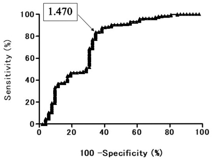
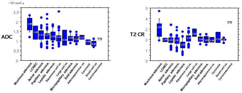
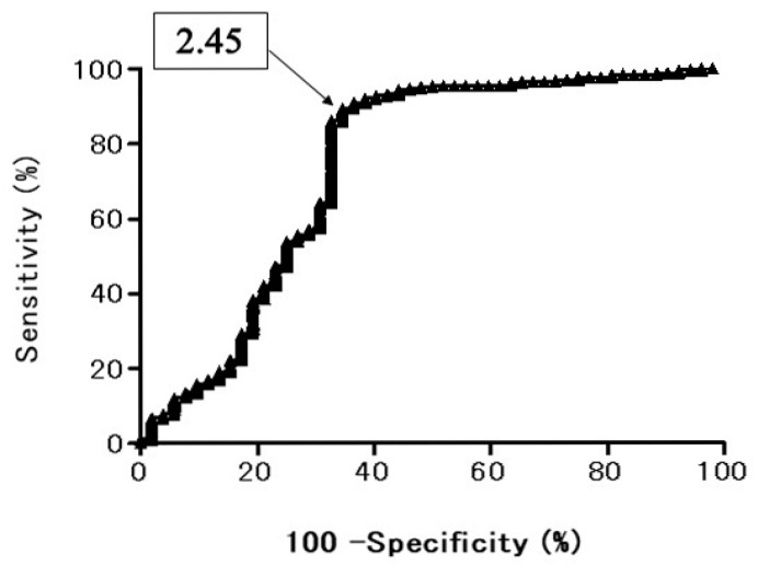
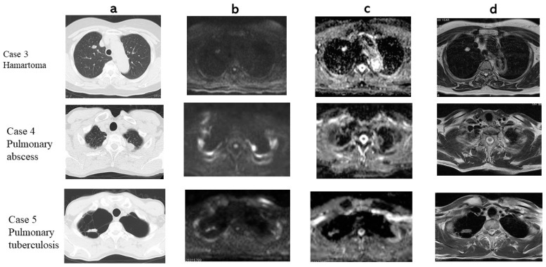
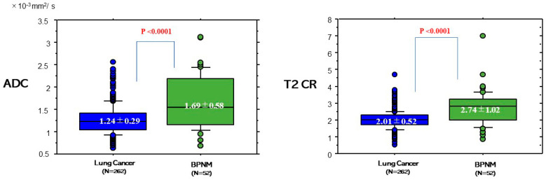
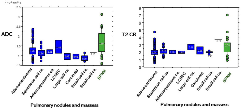
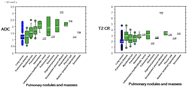
The purpose of this study is to determine whether the combination assessment of DWI and T2-weighted imaging (T2WI) improves the diagnostic ability for differential diagnosis of lung cancer from benign pulmonary nodules and masses (BPNMs). The optimal cut-off value (OCV) for differential diagnosis was set at 1.470 × 10 mm/s for apparent diffusion coefficient (ADC), and at 2.45 for T2 contrast ratio (T2 CR). The ADC (1.24 ± 0.29 × 10 mm/s) of lung cancer was significantly lower than that (1.69 ± 0.58 × 10 mm/s) of BPNM. The T2 CR (2.01 ± 0.52) of lung cancer was significantly lower than that (2.74 ± 1.02) of BPNM. As using the OCV for ADC, the sensitivity was 83.9% (220/262), the specificity 63.4% (33/52), and the accuracy 80.6% (253/314). As using the OCV for T2 CR, the sensitivity was 89.7% (235/262), the specificity 61.5% (32/52), and the accuracy 85.0% (267/314). In 212 PNMs which were judged to be malignant by both DWI and T2WI, 203 PNMs (95.8%) were lung cancers. In 33 PNMs which were judged to be benign by both DWI and T2WI, 23 PNMs (69.7%) were BPNMs. The combined assessment of DWI and T2WI could judge PNMs more precisely and would be acceptable for differential diagnosis of PNMs.

摘要

本研究的目的是确定弥散加权成像(DWI)与T2加权成像(T2WI)的联合评估是否能提高肺癌与良性肺结节及肿块(BPNM)鉴别诊断的诊断能力。鉴别诊断的最佳截断值(OCV)设定为表观扩散系数(ADC)为1.470×10⁻³mm²/s,T2对比率(T2 CR)为2.45。肺癌的ADC(1.24±0.29×10⁻³mm²/s)显著低于BPNM的ADC(1.69±0.58×10⁻³mm²/s)。肺癌的T2 CR(2.01±0.52)显著低于BPNM的T2 CR(2.74±1.02)。以ADC的OCV进行判断时,敏感性为83.9%(220/262),特异性为63.4%(33/52),准确性为80.6%(253/314)。以T2 CR的OCV进行判断时,敏感性为89.7%(235/262),特异性为61.5%(32/52),准确性为85.0%(267/314)。在DWI和T2WI均判定为恶性的212个肺结节中,203个(95.8%)为肺癌。在DWI和T2WI均判定为良性的33个肺结节中,23个(69.7%)为BPNM。DWI与T2WI的联合评估能更准确地判断肺结节,可用于肺结节的鉴别诊断。

https://cdn.ncbi.nlm.nih.gov/pmc/blobs/1ffb/8037373/fde9192e1896/cancers-13-01551-g008.jpg
https://cdn.ncbi.nlm.nih.gov/pmc/blobs/1ffb/8037373/72f99c595da7/cancers-13-01551-g001.jpg
https://cdn.ncbi.nlm.nih.gov/pmc/blobs/1ffb/8037373/06109383dc8b/cancers-13-01551-g002.jpg
https://cdn.ncbi.nlm.nih.gov/pmc/blobs/1ffb/8037373/f26aeb2ca8ae/cancers-13-01551-g003.jpg
https://cdn.ncbi.nlm.nih.gov/pmc/blobs/1ffb/8037373/4280ec1c7d16/cancers-13-01551-g004.jpg
https://cdn.ncbi.nlm.nih.gov/pmc/blobs/1ffb/8037373/439148ac7e29/cancers-13-01551-g005.jpg
https://cdn.ncbi.nlm.nih.gov/pmc/blobs/1ffb/8037373/447a193cd5f8/cancers-13-01551-g006.jpg
https://cdn.ncbi.nlm.nih.gov/pmc/blobs/1ffb/8037373/03730c530f83/cancers-13-01551-g007.jpg
https://cdn.ncbi.nlm.nih.gov/pmc/blobs/1ffb/8037373/fde9192e1896/cancers-13-01551-g008.jpg

相似文献

1
Combination Assessment of Diffusion-Weighted Imaging and T2-Weighted Imaging Is Acceptable for the Differential Diagnosis of Lung Cancer from Benign Pulmonary Nodules and Masses.扩散加权成像和T2加权成像联合评估可用于肺癌与良性肺结节及肿块的鉴别诊断。
Cancers (Basel). 2021 Mar 28;13(7):1551. doi: 10.3390/cancers13071551.
2
How to Discriminate Lung Cancer From Benign Pulmonary Nodules and Masses? Usefulness of Diffusion-Weighted Magnetic Resonance Imaging With Apparent Diffusion Coefficient and Inside/Wall Apparent Diffusion Coefficient Ratio.如何鉴别肺癌与良性肺结节及肿块?基于表观扩散系数和内部/壁表观扩散系数比值的扩散加权磁共振成像的效用
Clin Med Insights Oncol. 2021 Jul 7;15:11795549211014863. doi: 10.1177/11795549211014863. eCollection 2021.
3
Pulmonary Nodule and Mass: Superiority of MRI of Diffusion-Weighted Imaging and T2-Weighted Imaging to FDG-PET/CT.肺结节与肿块:扩散加权成像和T2加权成像的MRI相对于FDG-PET/CT的优势
Cancers (Basel). 2021 Oct 14;13(20):5166. doi: 10.3390/cancers13205166.
4
Novel Insights of T2-Weighted Imaging: Significance for Discriminating Lung Cancer from Benign Pulmonary Nodules and Masses.T2加权成像的新见解:对鉴别肺癌与良性肺结节及肿块的意义
Cancers (Basel). 2021 Jul 23;13(15):3713. doi: 10.3390/cancers13153713.
5
Diagnostic Usefulness of Combination of Diffusion-weighted Imaging and T2WI, Including Apparent Diffusion Coefficient in Breast Lesions: Assessment of Histologic Grade.弥散加权成像与 T2WI 联合应用,包括表观扩散系数在乳腺病变中的诊断价值:对组织学分级的评估。
Acad Radiol. 2018 May;25(5):643-652. doi: 10.1016/j.acra.2017.11.011. Epub 2018 Jan 12.
6
Free-breathing BLADE fat-suppressed T2 weighted turbo spin echo sequence for distinguishing lung cancer from benign pulmonary nodules or masses: A pilot study.自由呼吸的 BLADE 脂肪抑制 T2 加权涡轮自旋回波序列用于鉴别肺癌与良性肺结节或肿块:一项初步研究。
Magn Reson Imaging. 2023 Oct;102:79-85. doi: 10.1016/j.mri.2022.12.025. Epub 2023 Jan 2.
7
Differentiating benign and malignant inflammatory breast lesions: Value of T2 weighted and diffusion weighted MR images.鉴别良性和恶性炎性乳腺病变:T2加权和扩散加权磁共振成像的价值
Magn Reson Imaging. 2018 Jul;50:38-44. doi: 10.1016/j.mri.2018.03.012. Epub 2018 Mar 12.
8
Prostate cancer detection: comparison of T2-weighted imaging, diffusion-weighted imaging, proton magnetic resonance spectroscopic imaging, and the three techniques combined.前列腺癌检测:T2加权成像、扩散加权成像、质子磁共振波谱成像及三种技术联合应用的比较
Acta Radiol. 2008 Jun;49(5):602-10. doi: 10.1080/02841850802004983.
9
Diagnostic performance of diffusion weighted imaging of malignant and benign pulmonary nodules and masses: comparison with positron emission tomography.恶性与良性肺结节及肿块的扩散加权成像诊断效能:与正电子发射断层扫描的比较
Asian Pac J Cancer Prev. 2014;15(11):4629-35. doi: 10.7314/apjcp.2014.15.11.4629.
10
Diffusion-weighted MR Imaging and ADC Mapping in Differentiating Benign from Malignant Thyroid Nodules.扩散加权磁共振成像及表观扩散系数图在鉴别甲状腺良恶性结节中的应用
J Coll Physicians Surg Pak. 2015 Nov;25(11):785-8.

引用本文的文献

1
Zero-echo time magnetic resonance imaging for visualizing pulmonary neoplasms.用于可视化肺部肿瘤的零回波时间磁共振成像
BMC Med Imaging. 2025 Jul 7;25(1):274. doi: 10.1186/s12880-025-01666-5.
2
Diffusion-Weighted MRI: Potential Tool for Pulmonary Nodule Characterization.扩散加权磁共振成像:用于肺结节特征描述的潜在工具。
Indian J Radiol Imaging. 2023 Nov 23;34(1):1-2. doi: 10.1055/s-0043-1776884. eCollection 2024 Jan.
3
Predicting Non-Small-Cell Lung Cancer Survival after Curative Surgery via Deep Learning of Diffusion MRI.

本文引用的文献

1
Relationships and Qualitative Evaluation Between Diffusion-Weighted Imaging and Pathologic Findings of Resected Lung Cancers.肺癌切除标本的扩散加权成像与病理结果之间的关系及定性评估
Cancers (Basel). 2020 May 8;12(5):1194. doi: 10.3390/cancers12051194.
2
MRI in Evaluation of Solitary Pulmonary Nodules.磁共振成像在孤立性肺结节评估中的应用
Turk Thorac J. 2019 Jan 31;20(2):90-96. doi: 10.5152/TurkThoracJ.2018.18049. Print 2019 Apr.
3
Conventional MRI to detect the differences between mass-like tuberculosis and lung cancer.采用传统磁共振成像检测块状肺结核与肺癌之间的差异。
通过扩散磁共振成像的深度学习预测非小细胞肺癌根治性手术后的生存情况
Diagnostics (Basel). 2023 Aug 1;13(15):2555. doi: 10.3390/diagnostics13152555.
4
Differential value of diffusion kurtosis imaging and intravoxel incoherent motion in benign and malignant solitary pulmonary lesions.扩散峰度成像和体素内不相干运动在良恶性孤立性肺病变中的鉴别价值。
Front Oncol. 2023 Jan 12;12:1075072. doi: 10.3389/fonc.2022.1075072. eCollection 2022.
5
State of the Art: Lung Cancer Staging Using Updated Imaging Modalities.最新技术:使用更新的成像方式进行肺癌分期
Bioengineering (Basel). 2022 Sep 22;9(10):493. doi: 10.3390/bioengineering9100493.
6
Sensitivity and specificity of amide proton transfer-weighted imaging for assessing programmed death-ligand 1 status in non-small cell lung cancer: a comparative study with intravoxel incoherent motion and F-FDG PET.酰胺质子转移加权成像评估非小细胞肺癌程序性死亡配体1状态的敏感性和特异性:与体素内不相干运动及F-FDG PET的比较研究
Quant Imaging Med Surg. 2022 Sep;12(9):4474-4487. doi: 10.21037/qims-22-189.
7
Pulmonary Nodule and Mass: Superiority of MRI of Diffusion-Weighted Imaging and T2-Weighted Imaging to FDG-PET/CT.肺结节与肿块:扩散加权成像和T2加权成像的MRI相对于FDG-PET/CT的优势
Cancers (Basel). 2021 Oct 14;13(20):5166. doi: 10.3390/cancers13205166.
8
Automatic Detection of Colorectal Polyps Using Transfer Learning.基于迁移学习的结直肠息肉自动检测。
Sensors (Basel). 2021 Aug 24;21(17):5704. doi: 10.3390/s21175704.
9
Novel Insights of T2-Weighted Imaging: Significance for Discriminating Lung Cancer from Benign Pulmonary Nodules and Masses.T2加权成像的新见解:对鉴别肺癌与良性肺结节及肿块的意义
Cancers (Basel). 2021 Jul 23;13(15):3713. doi: 10.3390/cancers13153713.
J Thorac Dis. 2018 Oct;10(10):5673-5684. doi: 10.21037/jtd.2018.09.125.
4
Can Solitary Pulmonary Nodules Be Accurately Characterized with Diffusion-weighted MRI?扩散加权磁共振成像能否准确鉴别孤立性肺结节?
Radiology. 2019 Feb;290(2):535-536. doi: 10.1148/radiol.2018182442. Epub 2018 Nov 27.
5
Diagnostic Usefulness of Combination of Diffusion-weighted Imaging and T2WI, Including Apparent Diffusion Coefficient in Breast Lesions: Assessment of Histologic Grade.弥散加权成像与 T2WI 联合应用,包括表观扩散系数在乳腺病变中的诊断价值:对组织学分级的评估。
Acad Radiol. 2018 May;25(5):643-652. doi: 10.1016/j.acra.2017.11.011. Epub 2018 Jan 12.
6
Myometrial invasion and overall staging of endometrial carcinoma: assessment using fusion of T2-weighted magnetic resonance imaging and diffusion-weighted magnetic resonance imaging.子宫内膜癌的肌层浸润及整体分期:利用T2加权磁共振成像与扩散加权磁共振成像融合进行评估
Onco Targets Ther. 2017 Dec 15;10:5937-5943. doi: 10.2147/OTT.S145763. eCollection 2017.
7
The diagnostic accuracy of high b-value diffusion- and T-weighted imaging for the detection of prostate cancer: a meta-analysis.高 b 值弥散加权和 T 加权成像诊断前列腺癌的准确性:荟萃分析。
Abdom Radiol (NY). 2018 Jul;43(7):1787-1797. doi: 10.1007/s00261-017-1400-4.
8
Progressive massive fibrosis in patients with pneumoconiosis: utility of MRI in differentiating from lung cancer.尘肺病患者的进行性大块纤维化:MRI在与肺癌鉴别诊断中的应用
Acta Radiol. 2018 Jan;59(1):72-80. doi: 10.1177/0284185117700929. Epub 2017 Mar 31.
9
ADC Histogram Analysis of Cervical Cancer Aids Detecting Lymphatic Metastases-a Preliminary Study.ADC 直方图分析在宫颈癌淋巴结转移检测中的应用——一项初步研究。
Mol Imaging Biol. 2017 Dec;19(6):953-962. doi: 10.1007/s11307-017-1073-y.
10
Molecular magnetic resonance imaging in cancer.癌症中的分子磁共振成像
J Transl Med. 2015 Sep 23;13:313. doi: 10.1186/s12967-015-0659-x.