Seyed Jafari S Morteza, Blank Fabian, Ramser Hallie E, Woessner Alan E, Shafighi Maziar, Geiser Thomas, Quinn Kyle P, Hunger Robert E, Gazdhar Amiq
Department of Dermatology, Inselspital, Bern University Hospital, Bern, Switzerland.
Department for BioMedical Research (DBMR), University of Bern, Bern, Switzerland.
Front Surg. 2021 Mar 23;8:639661. doi: 10.3389/fsurg.2021.639661. eCollection 2021.
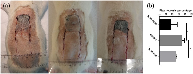
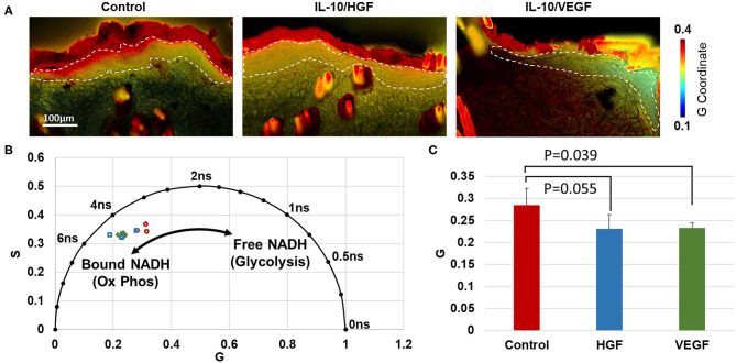
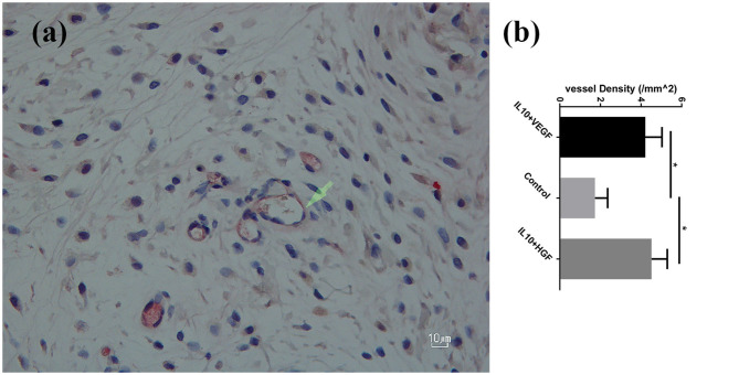
Preventing surgical flaps necrosis remains challenging. Laser Doppler imaging and ultrasound can monitor blood flow in flap regions, but they do not directly measure the cellular response to ischemia. The study aimed to investigate the efficacy of synergistic electroporation-mediated gene transfer of interleukin 10 (IL-10) with either hepatocyte growth factor (HGF) or vascular endothelial growth factor (VEGF) on the survival of a modified McFarlane flap, and to evaluate the effect of the treatment on cell metabolism, using label-free fluorescence lifetime imaging. Fifteen male Wistar rats (290-320 g) were randomly divided in three groups: group-A (control group) underwent surgery and received no gene transfer. Group-B received electroporation mediated hIL-10 gene delivery 24 h before and VEGF gene delivery 24 h after surgery. Group-C received electroporation mediated hIL-10 gene delivery 24 h before and hHGF gene delivery 24 h after surgery. The animals were assessed clinically and histologically. In addition, label-free fluorescence lifetime imaging was performed on the flap. Synergistic electroporation mediated gene delivery significantly decreased flap necrosis ( = 0.0079) and increased mean vessel density ( = 0.0079) in treatment groups B and C compared to control group-A. NADH fluorescence lifetime analysis indicated an increase in oxidative phosphorylation in the epidermis of the group-B ( = 0.039) relative to controls. These findings suggested synergistic electroporation-mediated gene transfer as a promising therapeutic approach to enhance viability and vascularity of skin flap. Furthermore, the study showed that combinational gene therapy promoted an increase in tissue perfusion and a relative increase in oxidative metabolism within the epithelium.
预防手术皮瓣坏死仍然具有挑战性。激光多普勒成像和超声可以监测皮瓣区域的血流,但它们不能直接测量细胞对缺血的反应。本研究旨在探讨白细胞介素10(IL-10)与肝细胞生长因子(HGF)或血管内皮生长因子(VEGF)协同电穿孔介导的基因转移对改良麦克法兰皮瓣存活的影响,并使用无标记荧光寿命成像评估该治疗对细胞代谢的作用。将15只雄性Wistar大鼠(290 - 320克)随机分为三组:A组(对照组)接受手术但未进行基因转移。B组在手术前24小时接受电穿孔介导的hIL-10基因递送,术后24小时接受VEGF基因递送。C组在手术前24小时接受电穿孔介导的hIL-10基因递送,术后24小时接受hHGF基因递送。对动物进行临床和组织学评估。此外,对皮瓣进行无标记荧光寿命成像。与A组对照组相比,B组和C组治疗组中协同电穿孔介导的基因递送显著降低了皮瓣坏死(P = 0.0079)并增加了平均血管密度(P = 0.0079)。NADH荧光寿命分析表明,相对于对照组,B组表皮中的氧化磷酸化增加(P = 0.039)。这些发现表明,协同电穿孔介导的基因转移是一种有前景的治疗方法,可提高皮瓣的活力和血管化。此外,该研究表明联合基因治疗促进了组织灌注的增加以及上皮内氧化代谢的相对增加。