Department of Plastic & Reconstructive Surgery, Shanghai Ninth People's Hospital, Shanghai Jiao Tong University School of Medicine, 639 Zhizhaoju Road, Shanghai, 200011, People's Republic of China.
Stem Cell Res Ther. 2021 Apr 15;12(1):243. doi: 10.1186/s13287-021-02318-5.
The regeneration response of the skin to mechanical stretching in vivo has been explored in reconstructive surgery to repair large-scale deformities. The ability of the skin to regenerate limits the reconstructive outcome. Here, we propose an approach in which autologous stromal vascular fraction (SVF) cells and mechanical stretching are combined to overcome this limitation and promote skin regeneration.
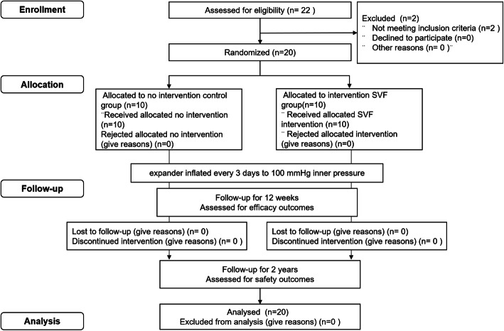
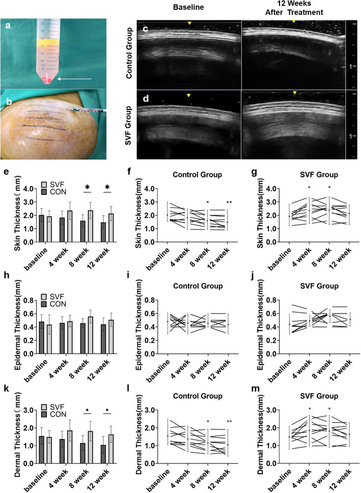
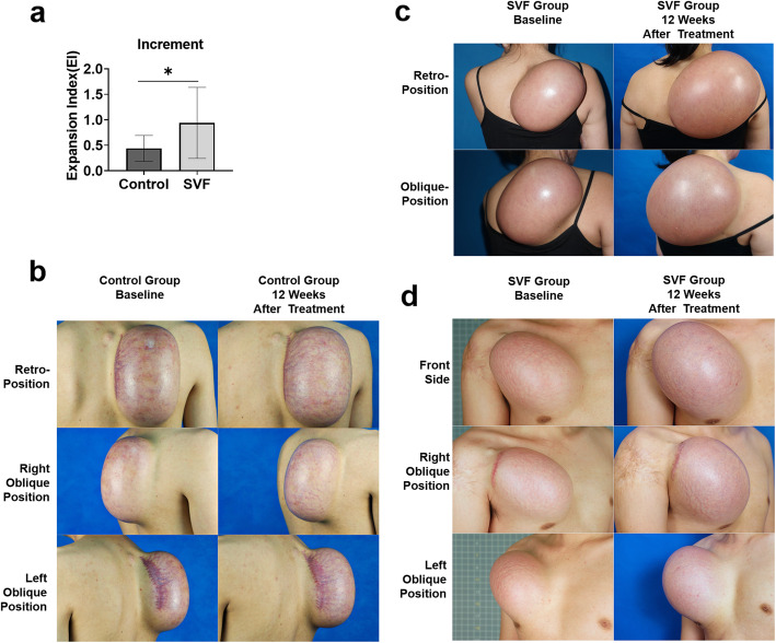
This randomized, blinded, placebo-controlled clinical trial screened 22 participants undergoing tissue expansion with exhausted regeneration. Twenty eligible participants received intradermal injections of the SVF or placebo treatments. Follow-ups were conducted at 4, 8, and 12 weeks to assess efficacy and at 2 years to assess safety. The primary endpoint was the expanded skin thickness at 12 weeks. The secondary endpoints included skin thickness at 4 and 8 weeks, the expansion index (EI), and the skin texture score at 12 weeks.
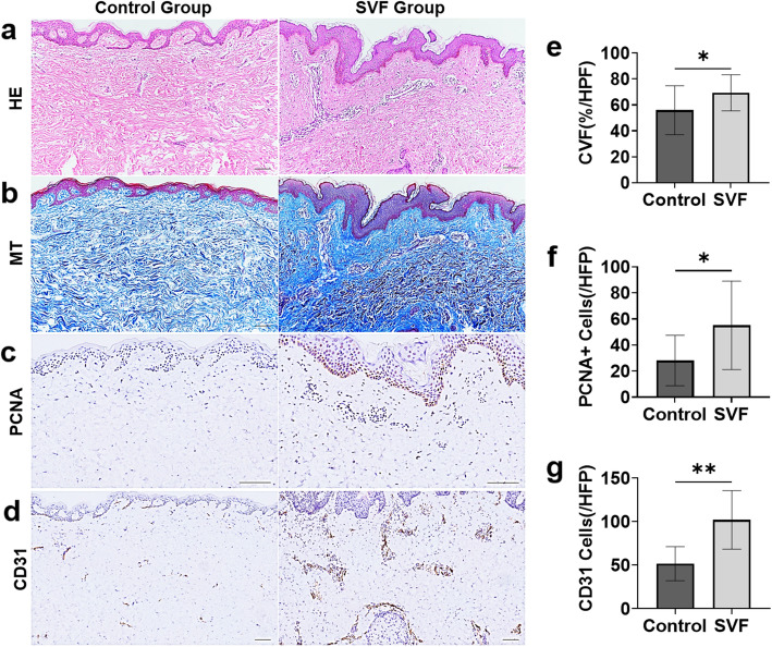
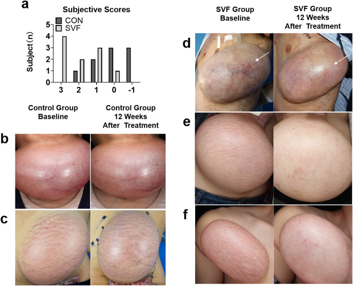
The skin thickness of the SVF group was significantly higher than that of the control group at both 8 weeks (mean difference 0.78 [95% CI - 1.43 to - 0.11]; p = 0.018) and 12 weeks (0.65 [95% CI - 1.30 to - 0.01]; p = 0.046). In the SVF group, the increase in skin thickness was significant at 4 weeks (0.49 [95% CI - 0.80 to - 0.06]; p = 0.010) to 8 weeks (0.45 [95% CI - 0.92 to 0.02]; p = 0.026) and maintained after 12 weeks, whereas that in the control group was reduced after 8 weeks (0.42 [95% CI - 0.07 to 0.91]; p = 0.037). The SVF group showed greater EI increases than the control group (0.50 [95% CI - 0.00 to 0.99]; p = 0.047). The skin texture scores in the SVF group were greater than those in the control group at 12 weeks. Histologically, SVF-treated expanded skin showed more proliferating cells and blood vessels, and the extracellular matrix volume increased. No severe adverse events occurred.
Transplantation of SVF cells can expedite the potency of mechanical stretch-induced skin regeneration and provide clinical reconstruction with plentiful tissue.
This trial was registered with the Chinese Clinical Trial Registry, ChiCTR2000039317 (registered 23 October 2020-retrospectively registered).
皮肤对体内机械拉伸的再生反应已在修复大面积畸形的重建外科中进行了探索。皮肤的再生能力限制了重建效果。在这里,我们提出了一种方法,即将自体基质血管成分 (SVF) 细胞与机械拉伸相结合,以克服这一限制并促进皮肤再生。
这是一项随机、盲法、安慰剂对照的临床试验,对 22 名接受组织扩张且再生能力耗尽的参与者进行了筛选。20 名符合条件的参与者接受了真皮内 SVF 或安慰剂治疗的注射。在 4、8 和 12 周时进行随访以评估疗效,并在 2 年时评估安全性。主要终点是 12 周时的扩张皮肤厚度。次要终点包括 4 周和 8 周时的皮肤厚度、扩张指数 (EI) 和 12 周时的皮肤质地评分。
SVF 组的皮肤厚度在 8 周时(平均差异 0.78 [95%CI -1.43 至 -0.11];p=0.018)和 12 周时(0.65 [95%CI -1.30 至 -0.01];p=0.046)均显著高于对照组。SVF 组在 4 周(0.49 [95%CI -0.80 至 -0.06];p=0.010)至 8 周(0.45 [95%CI -0.92 至 0.02];p=0.026)时皮肤厚度增加明显,并在 12 周后保持增加,而对照组在 8 周后减少(0.42 [95%CI -0.07 至 0.91];p=0.037)。SVF 组的 EI 增加大于对照组(0.50 [95%CI -0.00 至 0.99];p=0.047)。SVF 组的皮肤质地评分在 12 周时高于对照组。组织学检查显示,SVF 治疗的扩张皮肤显示出更多的增殖细胞和血管,细胞外基质体积增加。没有发生严重的不良事件。
SVF 细胞的移植可以加速机械拉伸诱导的皮肤再生能力,并为临床重建提供丰富的组织。
该试验在中国临床试验注册中心注册,ChiCTR2000039317(2020 年 10 月 23 日注册-回顾性注册)。