Laboratory of Membrane Protein Biology, National Centre for Cell Science, NCCS Complex, S. P. Pune University Campus, Ganeshkhind, Pune, Maharashtra, 411007, India.
The Maastricht Multimodal Molecular Imaging Institute (M4I), Division of Nanoscopy, Maastricht University, Maastricht, The Netherlands.
J Membr Biol. 2021 Jun;254(3):321-341. doi: 10.1007/s00232-021-00179-w. Epub 2021 May 5.
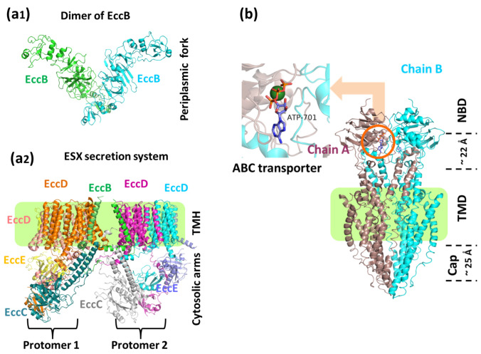
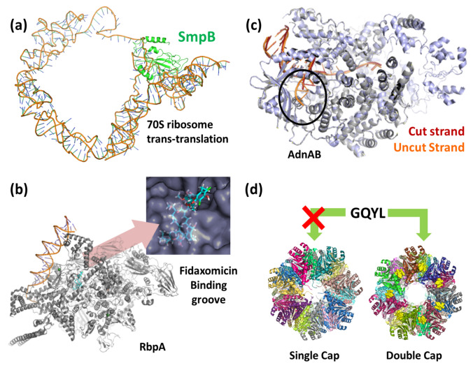
Mycobacterium tuberculosis (Mtb) is one of the deadliest pathogens encountered by humanity. Over the decades, its characteristic membrane organization and composition have been understood. However, there is still limited structural information and mechanistic understanding of the constituent membrane proteins critical for drug discovery pipelines. Recent advances in single-particle cryo-electron microscopy and cryo-electron tomography have provided the much-needed impetus towards structure determination of several vital Mtb membrane proteins whose structures were inaccessible via X-ray crystallography and NMR. Important insights into membrane composition and organization have been gained via a combination of electron tomography and biochemical and biophysical assays. In addition, till the time of writing this review, 75 new structures of various Mtb proteins have been reported via single-particle cryo-EM. The information obtained from these structures has improved our understanding of the mechanisms of action of these proteins and the physiological pathways they are associated with. These structures have opened avenues for structure-based drug design and vaccine discovery programs that might help achieve global-TB control. This review describes the structural features of selected membrane proteins (type VII secretion systems, Rv1819c, Arabinosyltransferase, Fatty Acid Synthase, F-type ATP synthase, respiratory supercomplex, ClpP1P2 protease, ClpB disaggregase and SAM riboswitch), their involvement in physiological pathways, and possible use as a drug target. Tuberculosis is a deadly disease caused by Mycobacterium tuberculosis. The Cryo-EM and tomography have simplified the understanding of the mycobacterial membrane organization. Some proteins are located in the plasma membrane; some span the entire envelope, while some, like MspA, are located in the mycomembrane. Cryo-EM has made the study of such membrane proteins feasible.
结核分枝杆菌(Mtb)是人类遇到的最致命病原体之一。几十年来,其特征性膜组织和组成已经被理解。然而,对于对于药物发现管道中关键的组成膜蛋白,仍然缺乏结构信息和机制理解。单颗粒冷冻电子显微镜和冷冻电子断层扫描的最新进展为结构测定提供了急需的动力,这些结构通过 X 射线晶体学和 NMR 无法获得。通过电子断层扫描和生化及生物物理测定的结合,获得了对膜组成和组织的重要见解。此外,在撰写本文时,通过单颗粒冷冻电子显微镜已经报道了 75 种不同 Mtb 蛋白的新结构。这些结构获得的信息提高了我们对这些蛋白作用机制以及它们所关联的生理途径的理解。这些结构为基于结构的药物设计和疫苗发现计划开辟了途径,这些计划可能有助于实现全球结核病控制。本文描述了选定膜蛋白(VII 型分泌系统、Rv1819c、阿拉伯糖基转移酶、脂肪酸合成酶、F 型 ATP 合酶、呼吸超级复合物、ClpP1P2 蛋白酶、ClpB 解聚酶和 SAM 核糖开关)的结构特征、它们在生理途径中的参与情况以及作为药物靶点的可能用途。结核病是由结核分枝杆菌引起的致命疾病。Cryo-EM 和断层扫描简化了对分枝杆菌膜组织的理解。一些蛋白位于质膜上;有些跨越整个包膜,而有些,如 MspA,则位于菌膜中。Cryo-EM 使得研究这些膜蛋白成为可能。