Hammed Ali, Mahfoud Moufid, Sulaiman Alaa, Najm Adnan, Hammed Salah
Tishreen University Hospital, Department of Neurosurgery, Lattakia, Syria.
Faculty of Medicine, Aleppo, Syria.
Ann Med Surg (Lond). 2021 Apr 21;65:102325. doi: 10.1016/j.amsu.2021.102325. eCollection 2021 May.
Meningiomas are extra-axial central nervous system (CNS) tumors that arise from the arachnoid cells of the dura mater. Only 1.8-3.2% of all meningiomas are located at foramen magnum (FM) and pure posterior FM meningioma are very rare. The diagnosis of malignancy in patients with meningiomas has been a controversial issue. Only a histological study can confirm this situation.
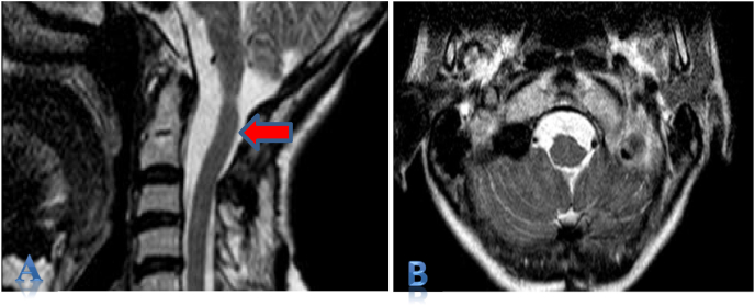
We report a case of A 52-year-old female presented with a history of neck pain with progressive spastic quadriparesis.
Magnetic resonance imaging MRI T1 and T2 weighted images revealed well-defied pure posterior foramen magnum Lesion. Although the lesion was very sticky to neurovascular components. Simpson grade I was achieved. Histopathology revealed Chordoid meningioma. The patient had a dramatic recovery.
Although choroid meningioma is usually well circumscribed, sticky tumors should be suspected. Recurrence of Chordoid meningioma should be suspected. Total excision should be achieved and routine follow-up should be informed. Reports about chordoid meningioma aren't common, but reports on choroid foramen magnum meningioma are very rare. The opportunity to give the patient a symptom-free and normal life should not be missed in such cases.
脑膜瘤是起源于硬脑膜蛛网膜细胞的轴外中枢神经系统(CNS)肿瘤。在所有脑膜瘤中,仅有1.8 - 3.2%位于枕骨大孔(FM),而单纯的枕骨大孔后部脑膜瘤非常罕见。脑膜瘤患者恶性肿瘤的诊断一直是一个有争议的问题。只有组织学研究才能证实这种情况。
我们报告一例52岁女性,有颈部疼痛伴进行性痉挛性四肢瘫病史。
磁共振成像(MRI)T1加权和T2加权图像显示边界清晰的单纯枕骨大孔后部病变。尽管该病变与神经血管成分粘连紧密,但仍实现了辛普森一级切除。组织病理学显示为脊索样脑膜瘤。患者恢复良好。
尽管脊索样脑膜瘤通常边界清晰,但对于粘连性肿瘤应予以怀疑。应怀疑脊索样脑膜瘤的复发。应实现全切并告知患者进行常规随访。关于脊索样脑膜瘤的报道并不常见,而关于枕骨大孔脊索样脑膜瘤的报道则极为罕见。在这种情况下,不应错过让患者过上无症状正常生活的机会。