Section of Nephrology, Department of Medicine, Boston University School of Medicine and Boston Medical Center, Boston, Massachussetts, USA; Renal Division, Brigham & Women's Hospital, Department of Medicine, Harvard Medical School, Boston, Massachussetts, USA.
Division of Nephrology, Department of Medicine, Kidney Research Institute, University of Washington, Seattle, Washington, USA.
Kidney Int. 2021 Sep;100(3):672-683. doi: 10.1016/j.kint.2021.04.037. Epub 2021 May 27.
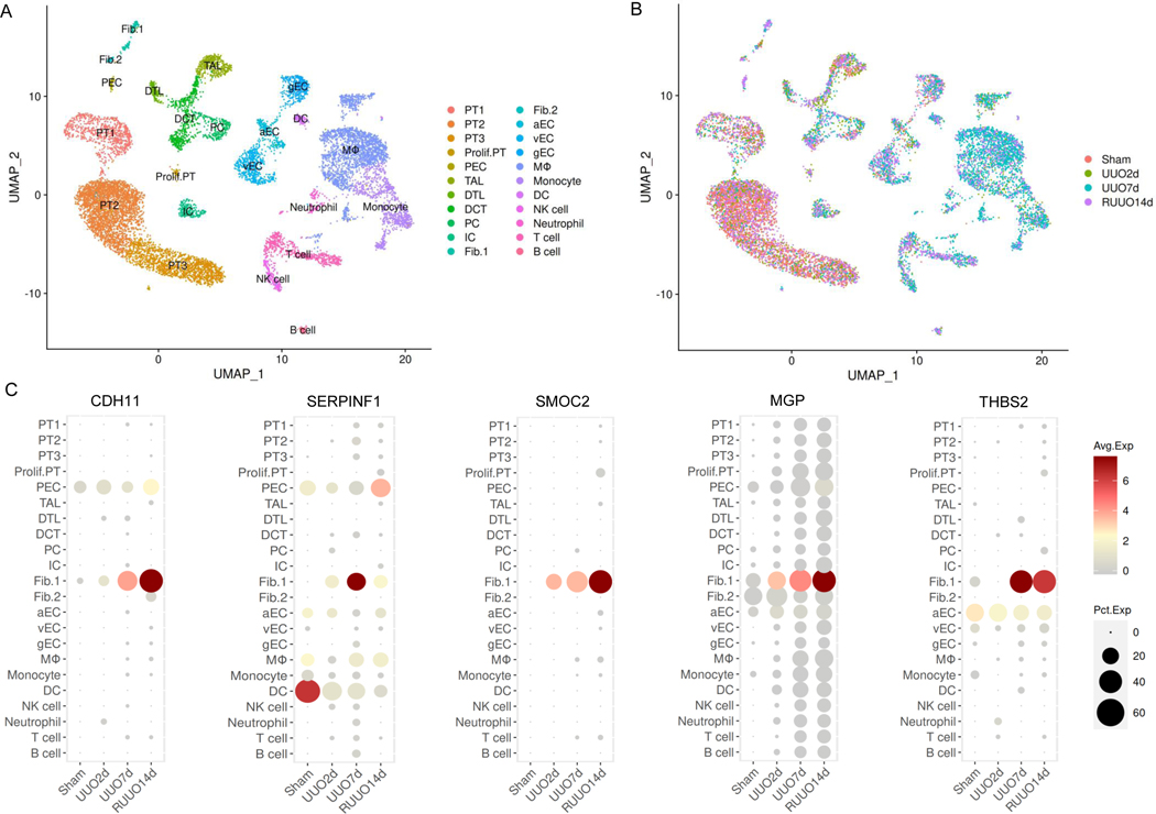
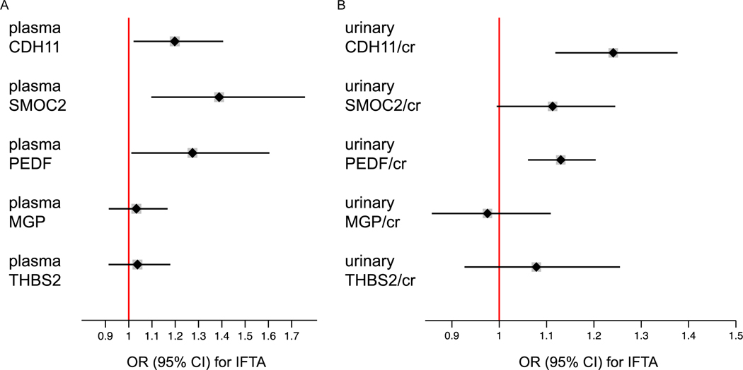
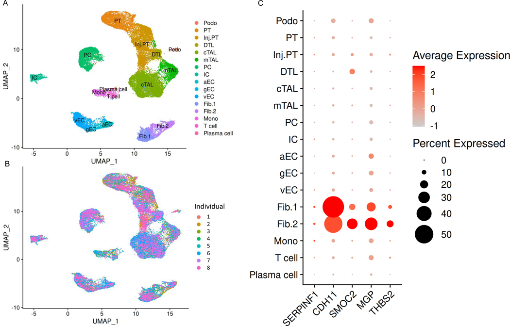
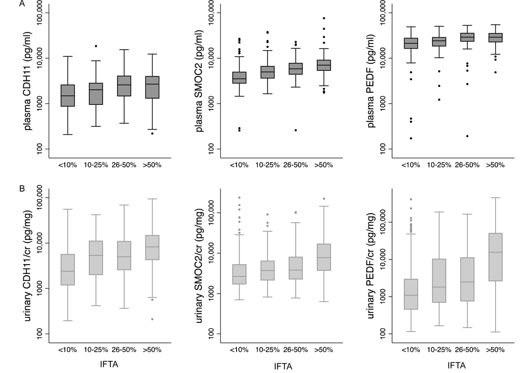
Kidney fibrosis constitutes the shared final pathway of nearly all chronic nephropathies, but biomarkers for the non-invasive assessment of kidney fibrosis are currently not available. To address this, we characterize five candidate biomarkers of kidney fibrosis: Cadherin-11 (CDH11), Sparc-related modular calcium binding protein-2 (SMOC2), Pigment epithelium-derived factor (PEDF), Matrix-Gla protein, and Thrombospondin-2. Gene expression profiles in single-cell and single-nucleus RNA-sequencing (sc/snRNA-seq) datasets from rodent models of fibrosis and human chronic kidney disease (CKD) were explored, and Luminex-based assays for each biomarker were developed. Plasma and urine biomarker levels were measured using independent prospective cohorts of CKD: the Boston Kidney Biopsy Cohort, a cohort of individuals with biopsy-confirmed semiquantitative assessment of kidney fibrosis, and the Seattle Kidney Study, a cohort of patients with common forms of CKD. Ordinal logistic regression and Cox proportional hazards regression models were used to test associations of biomarkers with interstitial fibrosis and tubular atrophy and progression to end-stage kidney disease and death, respectively. Sc/snRNA-seq data confirmed cell-specific expression of biomarker genes in fibroblasts. After multivariable adjustment, higher levels of plasma CDH11, SMOC2, and PEDF and urinary CDH11 and PEDF were significantly associated with increasing severity of interstitial fibrosis and tubular atrophy in the Boston Kidney Biopsy Cohort. In both cohorts, higher levels of plasma and urinary SMOC2 and urinary CDH11 were independently associated with progression to end-stage kidney disease. Higher levels of urinary PEDF associated with end-stage kidney disease in the Seattle Kidney Study, with a similar signal in the Boston Kidney Biopsy Cohort, although the latter narrowly missed statistical significance. Thus, we identified CDH11, SMOC2, and PEDF as promising non-invasive biomarkers of kidney fibrosis.
肾脏纤维化是几乎所有慢性肾病的共同终末途径,但目前尚无用于非侵入性评估肾脏纤维化的生物标志物。为了解决这个问题,我们鉴定了五个候选的肾脏纤维化生物标志物:钙黏蛋白 11(CDH11)、富含半胱氨酸的酸性分泌糖蛋白 2(SMOC2)、色素上皮衍生因子(PEDF)、基质 Gla 蛋白和血小板反应蛋白 2。在纤维化的啮齿动物模型和人类慢性肾病(CKD)的单细胞和单细胞核 RNA 测序(sc/snRNA-seq)数据集的单细胞和单细胞核 RNA 测序(sc/snRNA-seq)数据集中探索了这些候选生物标志物的基因表达谱,并为每个生物标志物开发了基于 Luminex 的检测方法。使用独立的 CKD 前瞻性队列测量了血浆和尿液生物标志物水平:波士顿肾脏活检队列,这是一个具有经活检确认的半定量肾脏纤维化评估的个体队列;以及西雅图肾脏研究队列,这是一个具有常见形式 CKD 的患者队列。有序逻辑回归和 Cox 比例风险回归模型用于检验生物标志物与间质纤维化和肾小管萎缩以及进展为终末期肾病和死亡的相关性。sc/snRNA-seq 数据证实了生物标志物基因在成纤维细胞中的细胞特异性表达。经过多变量调整后,在波士顿肾脏活检队列中,较高水平的血浆 CDH11、SMOC2 和 PEDF 以及尿液 CDH11 和 PEDF 与间质纤维化和肾小管萎缩的严重程度增加显著相关。在两个队列中,较高水平的血浆和尿液 SMOC2 和尿液 CDH11 与进展为终末期肾病独立相关。较高水平的尿液 PEDF 与西雅图肾脏研究中的终末期肾病相关,在波士顿肾脏活检队列中也有类似的信号,尽管后者略低于统计学意义。因此,我们确定 CDH11、SMOC2 和 PEDF 是有前途的肾脏纤维化非侵入性生物标志物。