Department of Medical Oncology, University Medical Center Groningen, University of Groningen, P.O. Box 30.001, 9700 RB, Groningen, The Netherlands.
Department of Nuclear Medicine and Molecular Imaging, University Medical Center Groningen, University Medical Center Groningen, Groningen, The Netherlands.
Eur J Nucl Med Mol Imaging. 2021 Dec;48(13):4369-4376. doi: 10.1007/s00259-021-05407-y. Epub 2021 Jun 2.
Immune checkpoint inhibitors can induce a T cell-mediated anti-tumor immune response in patients with melanoma. Visualizing T cell activity using positron emission tomography (PET) might allow early insight into treatment efficacy. Activated tumor-infiltrating T cells express the high-affinity interleukin-2 receptor (IL-2R). Therefore, we performed a pilot study, using fluorine-18-labeled IL-2 ([F]FB-IL2 PET), to evaluate whether a treatment-induced immune response can be detected.
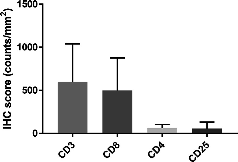
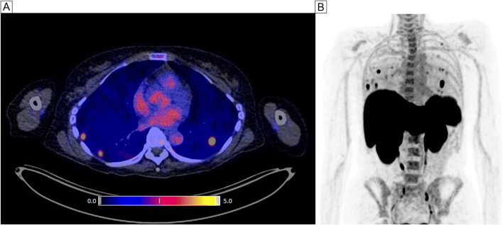
Patients with metastatic melanoma received ~ 200 MBq [F]FB-IL2 intravenously, followed by a PET/CT scan before and during immune checkpoint inhibitor therapy. [F]FB-IL2 uptake was measured as standardized uptake value in healthy tissues (SUV) and tumor lesions (SUV). Response to therapy was assessed using RECIST v1.1. Archival tumor tissues were used for immunohistochemical analyses of T cell infiltration.
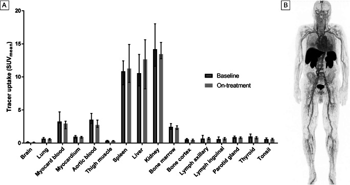
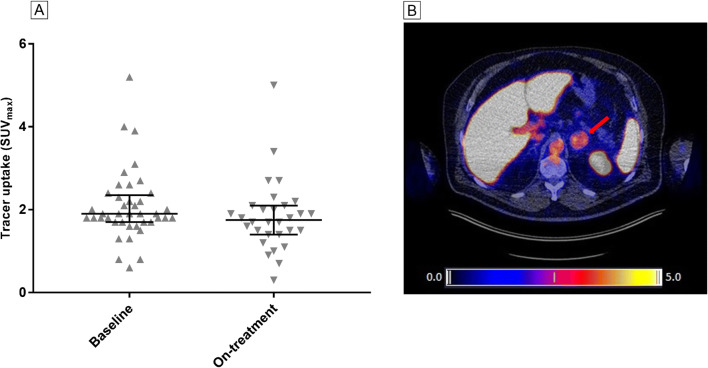
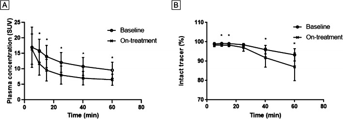
Baseline [F]FB-IL2 PET scans were performed in 13 patients. SUV at baseline was highest in the kidneys (14.2, IQR: 11.6-18.0) and liver (10.6, IQR: 8.6-13.4). In lymphoid tissues, uptake was highest in spleen (10.9, IQR: 8.8-12.4) and bone marrow (2.5, IQR: 2.1-3.0). SUV in tumor lesions (n = 41) at baseline was 1.9 (IQR: 1.7-2.3). In 11 patients, serial imaging was performed, three at week 6, seven at week 2, and one at week 4. Median [F]FB-IL2 tumor uptake decreased from 1.8 (IQR: 1.7-2.1) at baseline to 1.7 (IQR: 1.4-2.1) during treatment (p = 0.043). Changes in [F]FB-IL2 tumor uptake did not correlate with response. IL-2R expression in four archival tumor tissues was low and did not correlate with baseline [F]FB-IL2 uptake. No [F]FB-IL2-related side effects occurred.
PET imaging of the IL-2R, using [F]FB-IL2, is safe and feasible. In this small patient group, serial [F]FB-IL2-PET imaging did not detect a treatment-related immune response.
Clinicaltrials.gov : NCT02922283; EudraCT: 2014-003387.20.
免疫检查点抑制剂可在黑色素瘤患者中诱导 T 细胞介导的抗肿瘤免疫反应。使用正电子发射断层扫描(PET)可视化 T 细胞活性可能有助于早期了解治疗效果。活化的肿瘤浸润性 T 细胞表达高亲和力白细胞介素-2 受体(IL-2R)。因此,我们进行了一项初步研究,使用氟-18 标记的白细胞介素-2([F] FB-IL2 PET)来评估是否可以检测到治疗引起的免疫反应。
转移性黑色素瘤患者静脉注射约 200MBq [F] FB-IL2,然后在免疫检查点抑制剂治疗前和治疗期间进行 PET/CT 扫描。以健康组织(SUV)和肿瘤病变(SUV)的标准化摄取值测量 [F] FB-IL2 摄取。使用 RECIST v1.1 评估治疗反应。使用存档的肿瘤组织进行 T 细胞浸润的免疫组织化学分析。
13 名患者进行了基线 [F] FB-IL2 PET 扫描。基线时 SUV 最高的是肾脏(14.2,IQR:11.6-18.0)和肝脏(10.6,IQR:8.6-13.4)。在淋巴组织中,脾脏摄取量最高(10.9,IQR:8.8-12.4),骨髓摄取量最低(2.5,IQR:2.1-3.0)。基线时(n=41)肿瘤病变 SUV 为 1.9(IQR:1.7-2.3)。11 名患者进行了连续成像,3 名在第 6 周,7 名在第 2 周,1 名在第 4 周。中位[F] FB-IL2 肿瘤摄取从基线时的 1.8(IQR:1.7-2.1)降至治疗期间的 1.7(IQR:1.4-2.1)(p=0.043)。[F] FB-IL2 肿瘤摄取的变化与反应无关。四个存档肿瘤组织中的 IL-2R 表达较低,与基线 [F] FB-IL2 摄取无关。未发生与 [F] FB-IL2 相关的副作用。
使用 [F] FB-IL2 对 IL-2R 进行 PET 成像安全可行。在这个小患者组中,连续的[F] FB-IL2-PET 成像未检测到与治疗相关的免疫反应。
Clinicaltrials.gov:NCT02922283;EudraCT:2014-003387.20。