USC Epstein Family Center for Sports Medicine, Keck Medicine of USC, Los Angeles, CA, USA.
Department of Radiology, Keck School of Medicine of USC, Los Angeles, CA, USA.
Clin Orthop Surg. 2021 Jun;13(2):223-228. doi: 10.4055/cios20097. Epub 2021 Mar 9.
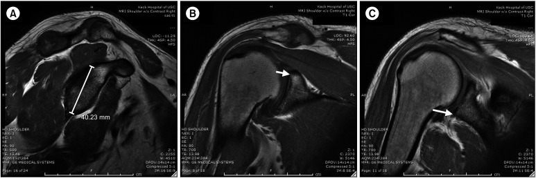
Recent literature suggests that three-dimensional magnetic resonance imaging (3D MRI) can replace 3D computed tomography (3D CT) when evaluating glenoid bone loss in patients with shoulder instability. We aimed to examine if 2D MRI in conjunction with a validated predictive formula for assessment of glenoid height is equivalent to the gold standard 3D CT scans for patients with recurrent glenohumeral instability.
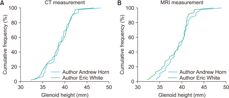
Patients with recurrent shoulder instability and available imaging were retrospectively reviewed. Glenoid height on 3D CT and 2D MRI was measured by two blinded raters. Difference and equivalence testing were performed using a paired -test and two one-sided tests, respectively. The interclass correlation coefficient (ICC) was used to test for interrater reliability, and percent agreement between the measurements of one reviewer was used to assess intrarater reliability.
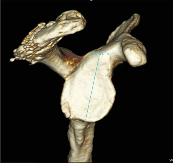
Using an equivalence margin of 1 mm, 3D CT and 2D MRI were found to be different ( = 0.123). The mean glenoid height was significantly different when measured on 2D MRI (39.09 ± 2.93 mm) compared to 3D CT (38.71 ± 2.89 mm) ( = 0.032). The mean glenoid width was significantly different between 3D CT (30.13 ± 2.43 mm) and 2D MRI (27.45 ± 1.72 mm) ( < 0.001). The 3D CT measurements had better interrater agreement (ICC, 0.91) than 2D MRI measurements (ICC, 0.8). intrarater agreement was also higher on CT.
Measurements of glenoid height using 3D CT and 2D MRI with subsequent calculation of the glenoid width using a validated methodology were not equivalent, and 3D CT was superior. Based on the validated methods for the measurement of glenoid bone loss on advanced imaging studies, 3D CT study must be preferred over 2D MRI in order to estimate the amount of glenoid bone loss in candidates for shoulder stabilization surgery and to assist in surgical decision-making.
最近的文献表明,在评估肩关节不稳定患者的肩盂骨丢失时,三维磁共振成像(3D MRI)可以替代三维计算机断层扫描(3D CT)。我们旨在研究二维 MRI 联合评估肩盂高度的验证预测公式是否与复发性肩盂不稳定患者的金标准 3D CT 扫描等效。
回顾性分析了患有复发性肩关节不稳定且有影像学资料的患者。由两名盲法评估者测量 3D CT 和 2D MRI 的肩盂高度。分别使用配对检验和两单边检验进行差异和等效性检验。采用组内相关系数(ICC)检验评估测量者间信度,采用一名评估者测量结果的百分比一致性检验评估测量者内信度。
使用 1mm 的等效边界,发现 3D CT 和 2D MRI 存在差异( = 0.123)。与 3D CT(38.71 ± 2.89mm)相比,2D MRI 上测量的肩盂高度明显不同(39.09 ± 2.93mm)( = 0.032)。3D CT(30.13 ± 2.43mm)与 2D MRI(27.45 ± 1.72mm)之间的肩盂宽度存在显著差异( < 0.001)。3D CT 测量的评估者间信度(ICC,0.91)优于 2D MRI 测量的评估者间信度(ICC,0.8)。CT 上的评估者内信度也更高。
使用 3D CT 和 2D MRI 测量肩盂高度,并随后使用经过验证的方法计算肩盂宽度,两种方法的测量结果并不等效,而且 3D CT 更优。基于先进影像学研究中肩盂骨丢失的测量的验证方法,为了评估肩稳定手术候选者的肩盂骨丢失量并辅助手术决策,必须首选 3D CT 研究,而不是 2D MRI。