Gupta Surendra Kumar, Nayak Nitish, Gandhoke Charandeep Singh, Gupta Rakesh Kumar, Sharma Anil Kumar, Singh Prashant, Sharma Raghvendra, Nehete Lokesh S
Department of Neurosurgery, AIIMS, Raipur, Chhattisgarh, India.
Department of Pathology and Laboratory Medicine, AIIMS, Raipur, Chhattisgarh, India.
Asian J Neurosurg. 2021 Feb 23;16(1):44-50. doi: 10.4103/ajns.AJNS_385_20. eCollection 2021 Jan-Mar.
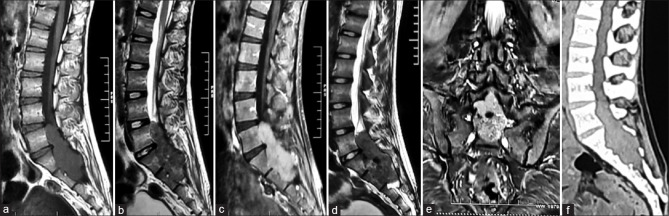
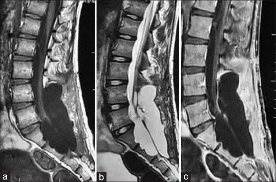
Spinal clear cell meningiomas (CCMs) are rare and dural-based lesion usually affecting the younger population. We report the rare case of giant nondural-based spinal CCM mimicking schwannoma and review the literature. A literature search was performed at PubMed and Embase until January 1, 2020. A total of 19 cases of nondural-based spinal CCM was reported. The following relevant data were extracted: authors, publication year, patient and tumor characteristics, treatment, and outcome. The mean age of the presentation was 20.58 years. Twelve (63.16%) were female and seven patients (36.84%) were male. The most common location was lumbosacral region 15 (79%). Fifteen (79%) tumors had cranio-caudal dimension ≤2 vertebral level, and only four (21%) tumors had dimension ≥2 vertebral level. Gross total resection (GTR) was performed in 18 (95%) patients and subtotal resection (STR) in 1 patient. Recurrences were reported in five (26.14%) patients. Four of them showed recurrences within 6 months; earliest at 2.3 months in the patient had undergone STR. Our patient is 19-year-old male diagnosed with a lumbosacral intradural lesion. Craniocaudal dimension is ≥2 vertebral level shows the foraminal extension and vertebral scalloping. GTR is performed. Intraoperatively, the tumor has foraminal extension and shows attachment with right S1S2 nerve root. No dural attachment is found. Six-month follow-up magnetic resonance image shows no evidence of disease. Nondural-based spinal CCMs are extremely rare and should be kept as a differential diagnosis in young patients with giant intradural tumor, and whose radiological features suggesting of schwannoma. It affects young patients and usually involves more than one vertebral level. The chances of recurrences and metastasis are always high even after GTR; hence, close follow-up of the entire neuraxis is warranted.
脊髓透明细胞脑膜瘤(CCMs)较为罕见,是一种起源于硬脑膜的病变,通常影响年轻人群。我们报告了一例罕见的巨大非硬脑膜起源的脊髓CCM,其表现类似神经鞘瘤,并对相关文献进行了综述。截至2020年1月1日,我们在PubMed和Embase上进行了文献检索。共报告了19例非硬脑膜起源的脊髓CCM。提取了以下相关数据:作者、发表年份、患者和肿瘤特征、治疗方法及结果。发病的平均年龄为20.58岁。女性12例(63.16%),男性7例(36.84%)。最常见的部位是腰骶部,共15例(79%)。15例(79%)肿瘤的头尾径≤2个椎体节段,只有4例(21%)肿瘤的头尾径≥2个椎体节段。18例(95%)患者接受了全切除(GTR),1例患者接受了次全切除(STR)。5例(26.14%)患者出现复发。其中4例在6个月内复发;接受STR的患者最早在2.3个月复发。我们的患者是一名19岁男性,被诊断为腰骶部硬膜内病变。头尾径≥2个椎体节段,显示有椎间孔扩展和椎体扇贝样改变。进行了GTR。术中,肿瘤有椎间孔扩展,与右侧S1S2神经根相连。未发现硬脑膜附着。6个月的随访磁共振成像显示无疾病迹象。非硬脑膜起源的脊髓CCM极为罕见,对于患有巨大硬膜内肿瘤且影像学特征提示神经鞘瘤的年轻患者,应将其作为鉴别诊断之一。它影响年轻患者,通常累及多个椎体节段。即使进行了GTR,复发和转移的几率也总是很高;因此,对整个神经轴进行密切随访是必要的。