• 文献检索
  • 文档翻译
  • 深度研究
  • 学术资讯
  • Suppr Zotero 插件Zotero 插件
  • 邀请有礼
  • 套餐&价格
  • 历史记录
应用&插件
Suppr Zotero 插件Zotero 插件浏览器插件Mac 客户端Windows 客户端微信小程序
定价
高级版会员购买积分包购买API积分包
服务
文献检索文档翻译深度研究API 文档MCP 服务
关于我们
关于 Suppr公司介绍联系我们用户协议隐私条款
关注我们

Suppr 超能文献

核心技术专利:CN118964589B侵权必究
粤ICP备2023148730 号-1Suppr @ 2026

文献检索

告别复杂PubMed语法,用中文像聊天一样搜索,搜遍4000万医学文献。AI智能推荐,让科研检索更轻松。

立即免费搜索

文件翻译

保留排版,准确专业,支持PDF/Word/PPT等文件格式,支持 12+语言互译。

免费翻译文档

深度研究

AI帮你快速写综述,25分钟生成高质量综述,智能提取关键信息,辅助科研写作。

立即免费体验

在选定的患者中,前额皮瓣移植1周后可完全切断蒂部。

Complete division of the pedicle of the forehead flap is possible after 1 week of engraftment in selected patients.

作者信息

Felcht Moritz, Wetzig Tino

机构信息

Department of Dermatology, Venereology and Allergy, Mannheim University Medical Center, Medical Faculty Mannheim, University of Heidelberg, Center of Excellence in Dermatology in Baden-Württemberg, European Center for Angioscience (ECAS), Medical Faculty Mannheim, University of Heidelberg, Mannheim, Germany.

Department of Dermatology, Dermatosurgery and Allergy, Asklepios Medical Center, Weissenfels, Germany.

出版信息

JAAD Int. 2020 Nov 30;2:5-11. doi: 10.1016/j.jdin.2020.10.004. eCollection 2021 Mar.

DOI:10.1016/j.jdin.2020.10.004
PMID:34409346
原文链接:https://pmc.ncbi.nlm.nih.gov/articles/PMC8361906/
Abstract

BACKGROUND

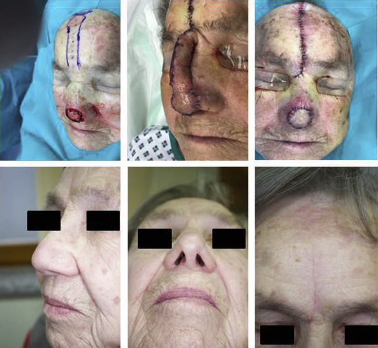
Recent studies have demonstrated that early division of the forehead flap (FHF) is possible if angiography is performed or a remnant of the pedicle is left behind. Whether or not careful selection of patients allows for complete division of the pedicle has not been studied.

OBJECTIVE

To assess if careful selection of patients allows for early complete division of the FHF.

METHODS

The exclusion criteria were trauma in the donor region, full-thickness defects, or a larger cartilage grafting. In the selected patients, complete division of the FHF pedicle was performed at early time points, when the pedicle was clinically engrafted (n = 12).

RESULTS

The median age of the patients was 80 years ± 8. The average size of the wounds was 6.6 cm ± 4.0. The complete division of the pedicle was performed in 10 patients after 7 days, 1 patient after 8 days, and 1 patient after 11 days (median 7.4 days ± 1.1). One patient developed a wound infection, and 1 suffered from postoperative bleeding. The latter patient was the only 1 who required debulking in a third surgical procedure. No necrosis or flap failures were observed.

LIMITATIONS

Retrospective, single-center study.

CONCLUSION

Careful selection allows for complete early division of the pedicle of FHF.

摘要

背景

近期研究表明,如果进行血管造影或保留蒂部残端,早期分割前额皮瓣(FHF)是可行的。但尚未研究过精心挑选患者是否能实现蒂部的完全分割。

目的

评估精心挑选患者是否能实现前额皮瓣的早期完全分割。

方法

排除标准为供区外伤、全层缺损或较大的软骨移植。在选定的患者中,当蒂部在临床上已成活时,于早期进行前额皮瓣蒂部的完全分割(n = 12)。

结果

患者的中位年龄为80岁±8岁。伤口平均大小为6.6 cm±4.0 cm。10例患者在7天后进行了蒂部的完全分割,1例在8天后,1例在11天后(中位时间7.4天±1.1天)。1例患者发生伤口感染,1例出现术后出血。后一名患者是唯一需要在第三次手术中进行减容的患者。未观察到坏死或皮瓣失败情况。

局限性

回顾性单中心研究。

结论

精心挑选患者可实现前额皮瓣蒂部的早期完全分割。

https://cdn.ncbi.nlm.nih.gov/pmc/blobs/f522/8361906/3713b4fb8a6d/gr3.jpg
https://cdn.ncbi.nlm.nih.gov/pmc/blobs/f522/8361906/22bb4cbd24cd/gr1.jpg
https://cdn.ncbi.nlm.nih.gov/pmc/blobs/f522/8361906/ff6b03ad2846/gr2.jpg
https://cdn.ncbi.nlm.nih.gov/pmc/blobs/f522/8361906/3713b4fb8a6d/gr3.jpg
https://cdn.ncbi.nlm.nih.gov/pmc/blobs/f522/8361906/22bb4cbd24cd/gr1.jpg
https://cdn.ncbi.nlm.nih.gov/pmc/blobs/f522/8361906/ff6b03ad2846/gr2.jpg
https://cdn.ncbi.nlm.nih.gov/pmc/blobs/f522/8361906/3713b4fb8a6d/gr3.jpg

相似文献

1
Complete division of the pedicle of the forehead flap is possible after 1 week of engraftment in selected patients.在选定的患者中,前额皮瓣移植1周后可完全切断蒂部。
JAAD Int. 2020 Nov 30;2:5-11. doi: 10.1016/j.jdin.2020.10.004. eCollection 2021 Mar.
2
Cost-effectiveness of Early Division of the Forehead Flap Pedicle.早期离断额部皮瓣蒂部的成本效益分析
JAMA Facial Plast Surg. 2017 Sep 1;19(5):418-420. doi: 10.1001/jamafacial.2017.0310.
3
Use of laser-assisted indocyanine green angiography for early division of the forehead flap pedicle.激光辅助吲哚菁绿血管造影术在前额皮瓣蒂早期离断中的应用。
JAMA Facial Plast Surg. 2015 May-Jun;17(3):209-14. doi: 10.1001/jamafacial.2015.0171.
4
Paramedian forehead flap pedicle division after 7 days using laser-assisted indocyanine green angiography.使用激光辅助吲哚菁绿血管造影术在7天后进行正中旁前额皮瓣蒂部离断。
J Plast Reconstr Aesthet Surg. 2021 Jan;74(1):116-122. doi: 10.1016/j.bjps.2020.05.079. Epub 2020 May 29.
5
A 10-year experience in nasal reconstruction with the three-stage forehead flap.应用三期前额皮瓣进行鼻再造的10年经验
Plast Reconstr Surg. 2002 May;109(6):1839-55; discussion 1856-61. doi: 10.1097/00006534-200205000-00010.
6
Reuse of the paramedian forehead flap pedicle: A case report.正中旁前额皮瓣蒂的再利用:一例报告。
SAGE Open Med Case Rep. 2023 Mar 21;11:2050313X231160913. doi: 10.1177/2050313X231160913. eCollection 2023.
7
The role of near-infrared fluorescence imaging with indocyanine green dye in pedicle division with the paramedian forehead flap.吲哚菁绿染料近红外荧光成像在额部中轴皮瓣皮瓣分割中的作用。
Int Wound J. 2021 Dec;18(6):881-888. doi: 10.1111/iwj.13590. Epub 2021 Mar 24.
8
A Cohort Study of Paramedian Forehead Flap in 2 Stages (87 Flaps) and 3 Stages (100 Flaps).两期(87例皮瓣)和三期(100例皮瓣)额旁正中皮瓣的队列研究。
Ann Plast Surg. 2015 Dec;75(6):615-9. doi: 10.1097/SAP.0000000000000209.
9
Paramedian forehead flap combined with hinge flap for nasal tip reconstruction.正中旁前额皮瓣联合铰链皮瓣用于鼻尖重建。
An Bras Dermatol. 2016 Sep-Oct;91(5 suppl 1):140-143. doi: 10.1590/abd1806-4841.20164583.
10
Medial femoral condyle free flap in combination with paramedian forehead flap for total/subtotal nasal reconstruction: Level of evidence: IV (therapeutic studies): Level of evidence: IV (therapeutic studies).股内侧髁游离皮瓣联合额正中皮瓣在全鼻/全鼻再造中的应用:证据等级:IV(治疗性研究):证据等级:IV(治疗性研究)。
Microsurgery. 2020 Mar;40(3):343-352. doi: 10.1002/micr.30532. Epub 2019 Nov 9.

引用本文的文献

1
Early Division of the Paramedian Forehead Flap: A Systematic Review and Retrospective Analysis.额旁正中皮瓣早期分叶:一项系统评价与回顾性分析
Laryngoscope. 2025 Jul;135(7):2233-2240. doi: 10.1002/lary.32009. Epub 2025 Jan 27.
2
Paramedian forehead flap survival after accidental flap transection on postoperative day 3.术后第3天意外切断皮瓣后正中旁前额皮瓣存活情况。
JAAD Case Rep. 2024 Jun 17;50:144-147. doi: 10.1016/j.jdcr.2024.05.039. eCollection 2024 Aug.

本文引用的文献

1
Reconstruction of Nasal Defects With Dermal Skin Substitutes-A Retrospective Study of 36 Defects.真皮替代物在鼻缺损重建中的应用——36 例缺损的回顾性研究。
J Cutan Med Surg. 2019 Jul/Aug;23(4):413-420. doi: 10.1177/1203475419852060. Epub 2019 Jun 10.
2
Postoperative Complications of Paramedian Forehead Flap Reconstruction.额部正中皮瓣重建术后并发症。
JAMA Facial Plast Surg. 2019 Jul 1;21(4):298-304. doi: 10.1001/jamafacial.2018.1855.
3
Reconstruction of defects of the proximal nasal sidewall using the procerus perforator flap.
J Dtsch Dermatol Ges. 2019 Feb;17(2):210-213. doi: 10.1111/ddg.13752.
4
Broadening applications and insights into the cross-paramedian forehead flap over a 19-year period.19 年来拓宽横额正中皮瓣的应用和认识。
J Plast Reconstr Aesthet Surg. 2019 May;72(5):763-770. doi: 10.1016/j.bjps.2018.12.001. Epub 2018 Dec 25.
5
Neovascularization Perfusion of Melolabial Flaps Using Intraoperative Indocyanine Green Angiography.术中吲哚菁绿血管造影评估穿支皮瓣的血管化及灌注。
JAMA Facial Plast Surg. 2019 May 1;21(3):230-236. doi: 10.1001/jamafacial.2018.1874.
6
Utility of Indocyanine Green Angiography to Identify Clinical Factors Associated With Perfusion of Paramedian Forehead Flaps During Nasal Reconstruction Surgery.吲哚菁绿血管造影在识别与鼻重建手术中额正中皮瓣灌注相关的临床因素中的应用。
JAMA Facial Plast Surg. 2019 May 1;21(3):206-212. doi: 10.1001/jamafacial.2018.1829.
7
The caudolaterally inserted transposition flap and its variations for reconstruction of defects of the nasal ala.用于重建鼻翼缺损的尾侧外侧转位皮瓣及其变异。
J Dtsch Dermatol Ges. 2017 Oct;15(10):981-987. doi: 10.1111/ddg.13318. Epub 2017 Aug 25.
8
[Single center analysis of the dermatosurgical patient cohort of a tumor center in Germany].[德国某肿瘤中心皮肤外科患者队列的单中心分析]
Hautarzt. 2017 May;68(5):377-384. doi: 10.1007/s00105-017-3951-2.
9
The Paramidline Forehead Flap: A Clinical and Microanatomic Study.
Dermatol Surg. 2016 Jun;42(6):764-71. doi: 10.1097/DSS.0000000000000722.
10
Use of laser-assisted indocyanine green angiography for early division of the forehead flap pedicle.激光辅助吲哚菁绿血管造影术在前额皮瓣蒂早期离断中的应用。
JAMA Facial Plast Surg. 2015 May-Jun;17(3):209-14. doi: 10.1001/jamafacial.2015.0171.