Zhang Jianping, Wang Lin, Xu Benhua, Huang Miaoyun, Chen Yuangui, Li Xiaobo
Department of Radiation Oncology, Fujian Medical University Union Hospital, Fuzhou, China.
Fujian Medical University Union Clinical Medicine College, Fujian Medical University, Fuzhou, China.
Front Oncol. 2021 Sep 16;11:705905. doi: 10.3389/fonc.2021.705905. eCollection 2021.
This study aimed to quantify the differences between pre- and post-contrast agent (CA) CT for CyberKnife brain SRS plans.
Twenty-five patients were retrospectively analyzed. They were divided into two categories, inhomogeneous cases (13 patients) and homogeneous cases (12 patients), according to whether the tumor was close to the cavity and inhomogeneous tissues or not. The pre-CA and post-CA plans were designed and calculated using the same monitor unit and paths as those in the ray-tracing algorithm, respectively.
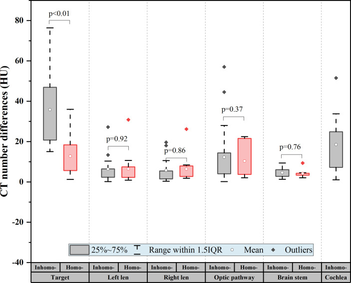
The CT number difference of tumor between pre- and post-CA was significant (on average, 24.78 ± 18.56 HU, -value < 0.01). The deviation value of the target was the largest at approximately 37 HU (inhomo-) and 13 HU (homo-) ( < 0.01), and the values of the organs at risk (OARs) were not statistically significant (-value > 0.05). However, it was not statistically significant for the dose difference between the two groups with the injection of CA (-value > 0.05). The absolute effective depth difference generally remained at a level of 1 mm, but the dose difference was quitely fluctuated sometimes more than 20%. The absolute effective depth difference of the inhomo-case (0.62 mm) was larger than that of the homo-case (0.37 mm) on median, as well as the variation amplitude (-value < 0.05). Moreover, the relative dose differences between the two cases were 0.38% (inhomo-) and 0.2% (homo-), respectively (-value < 0.05). At the criterion of 1 mm/1%, the gamma pass rate of the homo-case (95.89%) was larger than that of the inhomo-case (93.79%). For the OARs, except for the cochlea, the two cases were almost the same (>98.85%). The tumor control probability of the target was over 99.99% before and after injection of a CA, as well as the results for the homo-case and inhomo-case.
Considering the difference of evaluation indexes between pre- and post-CA images, we recommended plain CT to be employed as the primary image for improving the CK treatment accuracy of brain SRS, especially when the target was close to CA-sensitive OARs and cavity.
本研究旨在量化射波刀脑部立体定向放射治疗(SRS)计划中,注射对比剂(CA)前后CT图像的差异。
回顾性分析25例患者。根据肿瘤是否靠近腔隙及不均匀组织,将患者分为两类,即不均匀病例(13例)和均匀病例(12例)。分别使用与光线追踪算法相同的监测单位和路径,设计并计算注射对比剂前和注射对比剂后的计划。
注射对比剂前后肿瘤的CT值差异显著(平均为24.78±18.56HU,P值<0.01)。靶区偏差值在不均匀病例中约为37HU时最大,在均匀病例中约为13HU时最大(P<0.01),而危及器官(OARs)的值无统计学意义(P值>0.05)。然而,注射对比剂后两组间的剂量差异无统计学意义(P值>0.05)。绝对有效深度差异一般保持在1mm水平,但剂量差异有时波动较大,超过20%。不均匀病例的绝对有效深度差异中位数(0.62mm)大于均匀病例(0.37mm),变异幅度也是如此(P值<0.05)。此外,两种病例的相对剂量差异分别为0.38%(不均匀病例)和0.2%(均匀病例)(P值<0.05)。在1mm/1%的标准下,均匀病例的伽马通过率(95.89%)高于不均匀病例(93.79%)。对于危及器官,除耳蜗外,两种病例几乎相同(>98.85%)。注射对比剂前后靶区的肿瘤控制概率均超过99.99%,均匀病例和不均匀病例的结果也是如此。
考虑到注射对比剂前后图像评估指标的差异,我们建议采用平扫CT作为主要图像,以提高脑部SRS的射波刀治疗精度,尤其是当靶区靠近对对比剂敏感的危及器官和腔隙时。