Oral Radiology Area, Piracicaba Dental School, University of Campinas, Avenida Limeira, 901, Piracicaba, São Paulo, 13414-903, Brazil.
Oral Radiology Area, Dental School, Federal University of Mato Grosso do Sul, Campo Grande, MS, Brazil.
Oral Radiol. 2022 Oct;38(4):452-458. doi: 10.1007/s11282-021-00573-z. Epub 2021 Oct 9.
To evaluate the influence of the file format of digital periapical radiographs on the diagnosis of vertical root fracture (VRF).
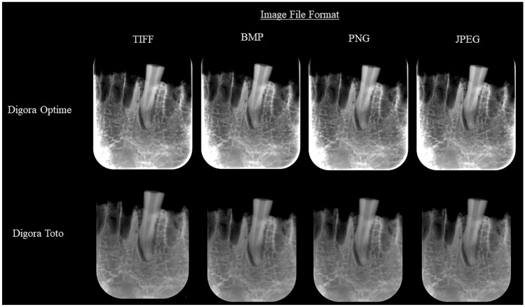
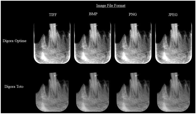
Periapical radiographic images of 34 single-rooted teeth-19 with VRF, and 15 without VRF were acquired using two digital systems-Digora Toto, and Digora Optime, and exported into four different file formats-TIFF, BMP, PNG, and JPEG, totaling 272 radiographs. The radiographs were assessed by five examiners for the detection of VRF, using a 5-point scale (1-definitely absent; 2-probably absent; 3-uncertain; 4-probably present; 5-definitely present). Diagnostic values of area under the ROC curve, specificity, and sensitivity for the diagnosis of VRF were calculated. The results were compared by two-way Analysis of Variance with post hoc Tukey's test. The intra- and inter-examiner agreements were measured by the Kappa test. The significance level was set at 5% for all analyses.
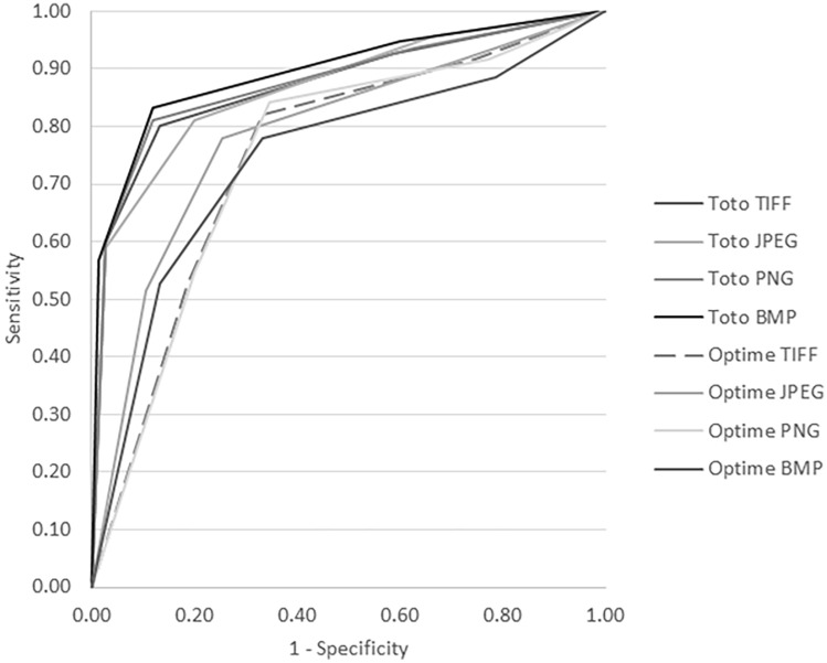
The values of intra-examiner agreement varied from moderate (0.56) to almost perfect (0.81), while the values of inter-examiner agreement varied from fair (0.29) to moderate (0.60). The image file format did not influence the diagnostic values for VRF for any of the radiographic systems tested (p > 0.05). Digora Toto had significantly greater values of area under the ROC curve than Digora Optime for all file formats (p = 0.001).
The image file format of periapical radiographs does not influence the diagnosis of VRF, regardless of the digital radiography system.
评估数字根尖片的文件格式对垂直根折(VRF)诊断的影响。
使用两种数字系统(Digora Toto 和 Digora Optime)获取 34 颗单根牙的根尖放射影像-19 颗有 VRF,15 颗无 VRF,并将其导出为四种不同的文件格式(TIFF、BMP、PNG 和 JPEG),共 272 张射线照片。由五名评估员使用 5 分制(1-肯定不存在;2-可能不存在;3-不确定;4-可能存在;5-肯定存在)评估这些射线照片以检测 VRF。计算了 ROC 曲线下面积、特异性和敏感性的诊断值,用于 VRF 的诊断。通过双向方差分析和事后 Tukey 检验比较结果。通过 Kappa 检验测量内部和外部评估者之间的一致性。所有分析的显着性水平均设置为 5%。
内部评估者的一致性值从中度(0.56)到几乎完美(0.81)不等,而外部评估者的一致性值从公平(0.29)到中度(0.60)不等。在所测试的任何放射系统中,图像文件格式均不影响 VRF 的诊断值(p>0.05)。对于所有文件格式,Digora Toto 的 ROC 曲线下面积值均显著大于 Digora Optime(p=0.001)。
无论使用哪种数字放射系统,根尖片的图像文件格式均不会影响 VRF 的诊断。