Department of Anesthesiology, Zhujiang Hospital, Southern Medical University, 253 Industrial Road, Guangzhou City, Guangdong Province, China.
Department of Laboratory Medicine, Zhujiang Hospital, Southern Medical University, 253 Industrial Road, Guangzhou City, Guangdong Province, China.
Oxid Med Cell Longev. 2021 Oct 7;2021:9925647. doi: 10.1155/2021/9925647. eCollection 2021.
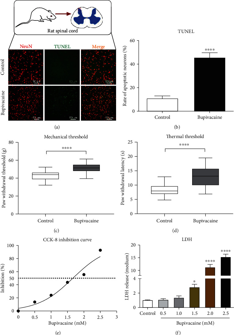
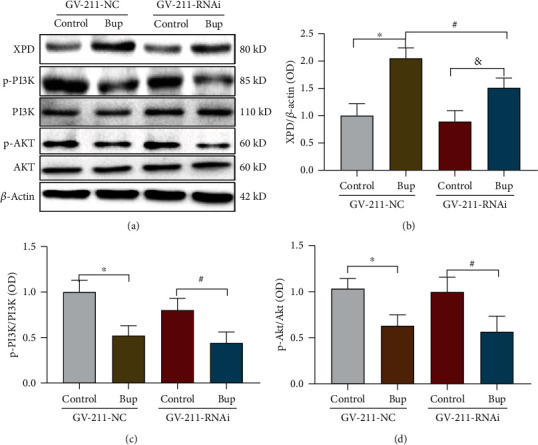
Bupivacaine, a local anesthetic widely used for regional anesthesia and pain management, has been reported to induce neuronal injury, especially DNA damage. Neurons employ different pathways to repair DNA damage. However, the mechanism underlying bupivacaine-mediated DNA damage repair is unclear. A rat neuronal injury model was established by intrathecal injection of (3%) bupivacaine. An in vitro neuronal injury model was generated by exposing SH-SY5Y cells to bupivacaine (1.5 mmol/L). Then, a cDNA plate array was used to identify the DNA repair genes after bupivacaine exposure. The results showed that xeroderma pigmentosum complementary group D (XPD) of the nuclear excision repair (NER) pathway was closely associated with the repair of DNA damage induced by bupivacaine. Subsequently, Western blot assay and immunohistochemistry indicated that the expression of the repair enzyme XPD was upregulated after DNA damage. Downregulation of XPD expression by a lentivirus aggravated the DNA damage induced by bupivacaine. In addition, phosphatidyl-3-kinase (PI3K)/AKT signaling in neurons was inhibited after exposure to bupivacaine. After PI3K/AKT signaling was inhibited, bupivacaine-mediated DNA damage was further aggravated, and the expression of XPD was further upregulated. However, knockdown of XPD aggravated bupivacaine-mediated neuronal injury but did not affect PI3K/AKT signaling. In conclusion, the repair enzyme XPD, which was partially regulated by PI3K/AKT signaling, responded to bupivacaine-mediated neuronal DNA damage. These results can be used as a reference for the treatment of bupivacaine-induced neurotoxicity.
布比卡因是一种广泛用于局部麻醉和疼痛管理的局麻药,已被报道可诱导神经元损伤,尤其是 DNA 损伤。神经元利用不同的途径来修复 DNA 损伤。然而,布比卡因介导的 DNA 损伤修复的机制尚不清楚。通过鞘内注射(3%)布比卡因建立大鼠神经元损伤模型。通过暴露于布比卡因(1.5mmol/L)来建立体外神经元损伤模型。然后,使用 cDNA 板阵列来鉴定布比卡因暴露后与 DNA 修复相关的基因。结果表明,核切除修复(NER)途径中的着色性干皮病互补组 D(XPD)与布比卡因诱导的 DNA 损伤修复密切相关。随后,Western blot 检测和免疫组织化学分析表明,修复酶 XPD 的表达在 DNA 损伤后上调。通过慢病毒下调 XPD 表达加重了布比卡因诱导的 DNA 损伤。此外,暴露于布比卡因后,神经元中的磷酸肌醇 3-激酶(PI3K)/AKT 信号被抑制。PI3K/AKT 信号被抑制后,布比卡因介导的 DNA 损伤进一步加重,XPD 的表达进一步上调。然而,XPD 的敲低加重了布比卡因介导的神经元损伤,但不影响 PI3K/AKT 信号。总之,部分受 PI3K/AKT 信号调节的修复酶 XPD 对布比卡因介导的神经元 DNA 损伤做出反应。这些结果可作为治疗布比卡因诱导的神经毒性的参考。