Zhang Qi-Chen, Zou Yan-Pei, Hu Shun-Qi, Zhang Tai-Wei, Zhou Hao, Liang Bing, Zhuang Chen-Yang, Wang Hui-Ren, Jiang Li-Bo, Li Xi-Lei
Department of Orthopaedic Surgery, Zhongshan Hospital, Fudan University, Shanghai, China.
Department of Orthopedic Surgery, Zhongshan Hospital, Fudan University (Xiamen Branch), Xiamen, China.
Ann Transl Med. 2021 Sep;9(17):1376. doi: 10.21037/atm-21-227.
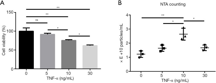
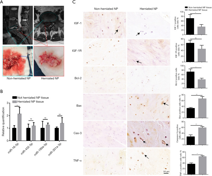
Exosomes may contain excess cellular components released by cells in response to harmful external stimuli to maintain cellular homeostasis. Inflammatory cytokines, such as tumor necrosis factor-alpha (TNF-α), can induce cell apoptosis, alter cellular component expression levels, and stimulate exosome release. In this study, we examined whether exosomes released from nucleus pulposus cells (NPCs) under inflammatory conditions could induce normal NP cell apoptosis in rats and its underlining mechanism.
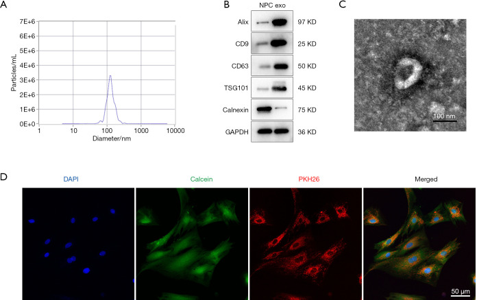
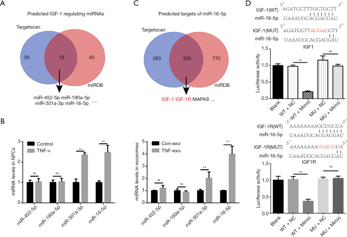
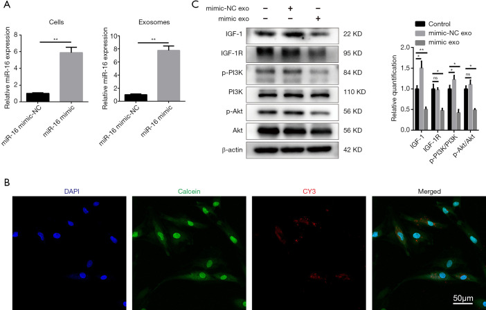
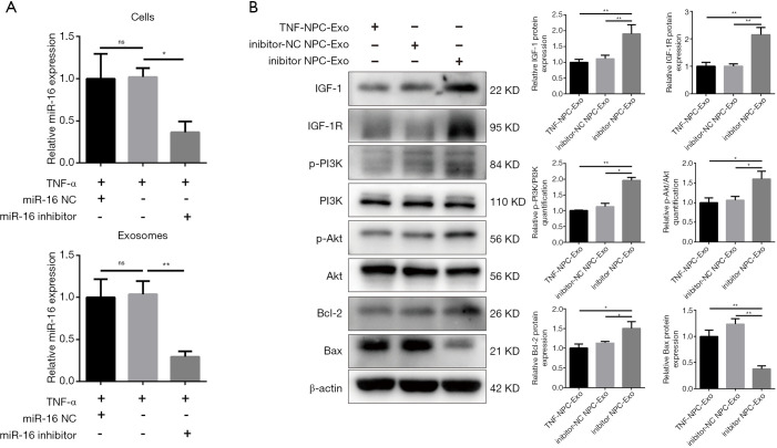
Exosomes were isolated from TNF-α-treated NPCs and used to treat normal NPCs. The effects were assessed by flow cytometry and western blot analysis. Anti-apoptotic insulin-like growth factor-1 (IGF-1) expression in NPCs was assessed by western blot analysis. Given the exosomal miRNAs might be the key factors of exosomes, bioinformatics approaches and quantitative real-time polymerase chain reaction (qRT-PCR) were used to identify IGF-1-regulating micro RNAs (miRNAs), including miR-16. Luciferase reporter assay assessed miR-16 regulation of IGF-1 and IGF-1 receptor (IGF-1R). NPCs were transfected with miR-16 mimic, and exosomes were applied to normal NPCs. NPCs were pretreated with 10 ng/mL TNF-α, transfected with miR-16 inhibitors, and the exosomes were isolated. Cell and exosome miR-16 levels were detected by qRT-PCR. Western blot analysis determined IGF-1, IGF-1R, and apoptotic marker levels in exosome-treated NPCs.
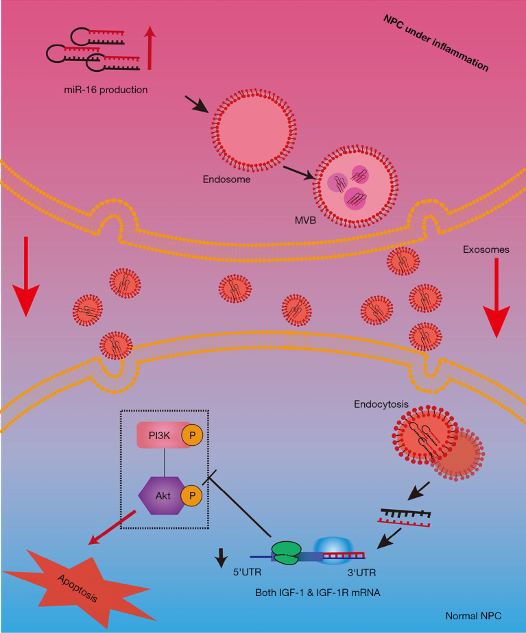
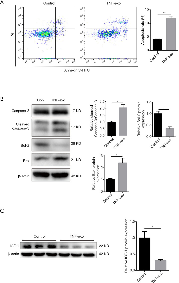
Exosomes from TNF-α-treated NPCs induced apoptosis in normal NPCs and repressed IGF-1 expression. Exosomal miR-16 regulated IGF-1 and induced NPC apoptosis. The dual-luciferase reporter assay revealed that miR-16 binds the 3' untranslated regions (3'-UTRs) of IGF-1 and IGF-1R. Exosomal miR-16 repressed IGF-1 and the IGF-1R/phosphoinositide 3-kinase (PI3K)/protein kinase B (Akt) pathway which therefore induced NPC apoptosis. Rescue experiments using miR-16 inhibitors further validated these findings.
The inflammatory factor TNF-α stimulated exosome release from NPCs, which induced the apoptosis of normal NPCs through the actions of exosomal miR-16. Exosomal miR-16 directly repressed the anti-apoptotic IGF-1/IGF-1R pathway, increasing the apoptosis of NPCs.
外泌体可能包含细胞在应对有害外部刺激时释放的多余细胞成分,以维持细胞内稳态。炎性细胞因子,如肿瘤坏死因子-α(TNF-α),可诱导细胞凋亡、改变细胞成分表达水平并刺激外泌体释放。在本研究中,我们检测了炎性条件下髓核细胞(NPCs)释放的外泌体是否能诱导大鼠正常NP细胞凋亡及其潜在机制。
从TNF-α处理的NPCs中分离出外泌体,并用于处理正常NPCs。通过流式细胞术和蛋白质印迹分析评估效果。通过蛋白质印迹分析评估NPCs中抗凋亡胰岛素样生长因子-1(IGF-1)的表达。鉴于外泌体微小RNA(miRNAs)可能是外泌体的关键因素,采用生物信息学方法和定量实时聚合酶链反应(qRT-PCR)来鉴定调节IGF-1的微小RNA(miRNAs),包括miR-16。荧光素酶报告基因检测评估miR-16对IGF-1和IGF-1受体(IGF-1R)的调节作用。用miR-16模拟物转染NPCs,并将外泌体应用于正常NPCs。用10 ng/mL TNF-α预处理NPCs,转染miR-16抑制剂,然后分离外泌体。通过qRT-PCR检测细胞和外泌体miR-16水平。蛋白质印迹分析测定外泌体处理的NPCs中IGF-1、IGF-1R和凋亡标志物水平。
TNF-α处理的NPCs释放的外泌体诱导正常NPCs凋亡并抑制IGF-1表达。外泌体miR-16调节IGF-1并诱导NPC凋亡。双荧光素酶报告基因检测显示miR-16与IGF-1和IGF-1R的3'非翻译区(3'-UTRs)结合。外泌体miR-16抑制IGF-1和IGF-1R/磷脂酰肌醇3-激酶(PI3K)/蛋白激酶B(Akt)通路,从而诱导NPC凋亡。使用miR-16抑制剂的挽救实验进一步验证了这些发现。
炎性因子TNF-α刺激NPCs释放外泌体,外泌体通过外泌体miR-16的作用诱导正常NPCs凋亡。外泌体miR-16直接抑制抗凋亡的IGF-1/IGF-1R通路,增加NPCs的凋亡。