Liu Tang, Hu Jiawei, Liu Yajie, Chen Honghai, Guo Dongmei
Department of Radiology, The Second Affiliated Hospital of Dalian Medical University, Dalian, China.
Ann Transl Med. 2021 Oct;9(20):1569. doi: 10.21037/atm-21-4884.
This study aimed to evaluate the diagnostic accuracy of diffusion kurtosis imaging (DKI) in differentiating early hepatic fibrosis (HF) from normal liver and advanced HF in rabbits.
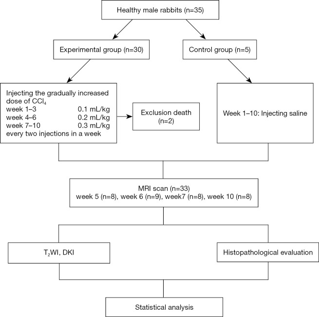
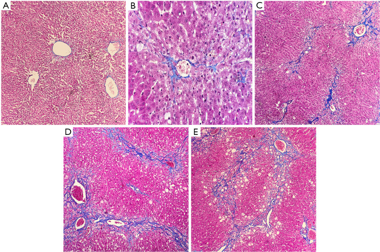
A total of 35 healthy New Zealand white rabbits were included in the study. A model of HF was established in 30 rabbits through subcutaneous injections of 50% carbon tetrachloride (CCl)/olive oil, while 5 rabbits received saline injections. The gradually increased doses of CCl were 0.1, 0.2, and 0.3 mL/kg in weeks 1 to 3, weeks 4 to 6, and weeks 7 to 10, respectively. Two injections were given each week. Two rabbits in the experimental group died. All rabbits underwent DKI with three b values (0, 500, and 1,000 s/mm) at week 5 (n=8), week 6 (n=9), week 7 (n=8), and week 10 (n=8). Approximately 2 liver lobes per rabbit were selected for histopathology. Mean diffusivity (MD) and mean kurtosis (MK) were calculated. Discrimination capacities of DKI parameters were analyzed and compared by receiver operating characteristic (ROC) analysis.
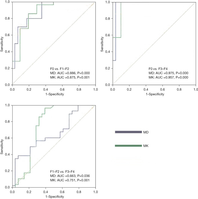
The meta-analysis of histological data in viral hepatitis (METAVIR) scoring system was used to classify liver lobes into the control group (F0, n=0), early HF group (F1-F2, n=28), and advanced HF group (F3-F4, n=28). MD and MK values were significantly different among the three groups (all P<0.05). MD value was negatively correlated with increased fibrosis level, while MK value was positively correlated with increased fibrosis level (ρ=-0.540, 0.614; P<0.05). The area under ROC curves (AUCs) for MD and MK were 0.886 and 0.875, respectively, for characterization of F0 and F1-F2, and 0.975 and 0.957 for F0 and F3-F4. AUC for MK was 0.751 for characterization of F1-F2 and F3-F4. MD performed better than MK for characterization of F0 and F1-F2 as well as F0 and F3-F4. MK showed good differentiation performance between F1-F2 and F3-F4.
Our results showed that DKI contributed to discriminating reversible early HF from normal liver and advanced HF and as a result, showed promise for use in HF diagnosis.
本研究旨在评估扩散峰度成像(DKI)在区分兔早期肝纤维化(HF)与正常肝脏及晚期HF方面的诊断准确性。
本研究共纳入35只健康的新西兰白兔。30只兔子通过皮下注射50%四氯化碳(CCl)/橄榄油建立HF模型,5只兔子注射生理盐水。在第1至3周、第4至6周和第7至10周,CCl的注射剂量逐渐增加,分别为0.1、0.2和0.3 mL/kg。每周注射两次。实验组有两只兔子死亡。所有兔子在第5周(n = 8)、第6周(n = 9)、第7周(n = 8)和第10周(n = 8)接受了具有三个b值(0、500和1000 s/mm²)的DKI检查。每只兔子选取约2个肝叶进行组织病理学检查。计算平均扩散率(MD)和平均峰度(MK)。通过受试者操作特征(ROC)分析对DKI参数的鉴别能力进行分析和比较。
采用病毒性肝炎组织学数据的荟萃分析(METAVIR)评分系统将肝叶分为对照组(F0,n = 0)、早期HF组(F1 - F2,n = 28)和晚期HF组(F3 - F4,n = 28)。三组间MD和MK值差异有统计学意义(均P < 0.05)。MD值与纤维化程度增加呈负相关;而MK值与纤维化程度增加呈正相关(ρ = -0.540,0.614;P < 0.05)。对于区分F0和F1 - F2,MD和MK的ROC曲线下面积(AUC)分别为为0.886和0.875,对于区分F0和F3 - F4,AUC分别为0.975和0.957。对于区分F1 - F2和F3 - F4,MK的AUC为0.751。在区分F0和F1 - F2以及F0和F3 - F4方面,MD的表现优于MK。MK在区分F1 - F2和F3 - F4方面表现出良好的鉴别性能。
我们的结果表明,DKI有助于区分可逆性早期HF与正常肝脏及晚期HF,因此在HF诊断中具有应用前景。