Yale University School of Medicine, PO Box 208042, New Haven, CT, 06520-8042, USA.
Quinnipiac University, 275 Mount Carmel Ave, CT, 06518, Hamden, USA.
Childs Nerv Syst. 2022 Feb;38(2):287-294. doi: 10.1007/s00381-021-05408-0. Epub 2021 Nov 23.
An extensive literature has postulated multiple etiologies for aqueductal stenosis. No publications were found, discussing that evolutionary modifications might explain aqueductal anomalies. This study's objectives were to review the evolutionary modifications of vertebrates' tectum structures that might explain human aqueduct anomalies. Undertaking vertebrate comparative study is currently not feasible in view of limitations in obtaining vertebrate material. Thus, vertebrate material collected, injected, dissected, and radiographed in the early 1970s was analyzed, focusing on the aqueduct and components of the midbrain tectum.
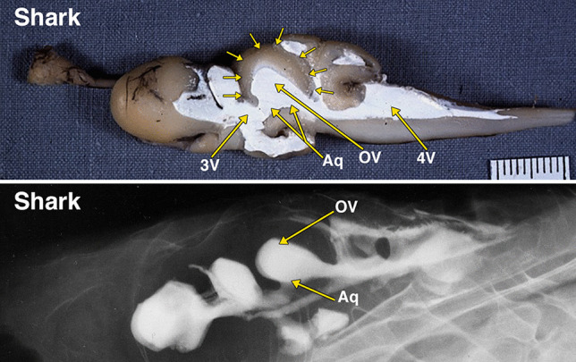
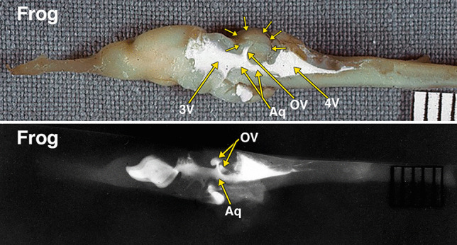
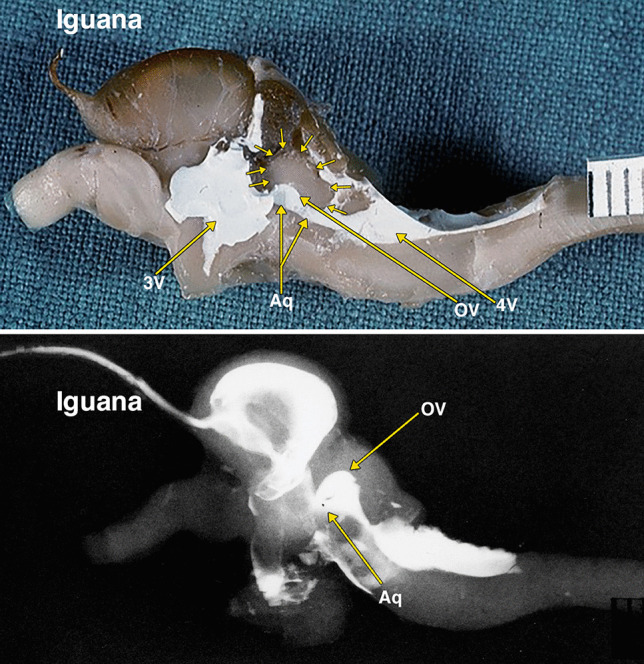
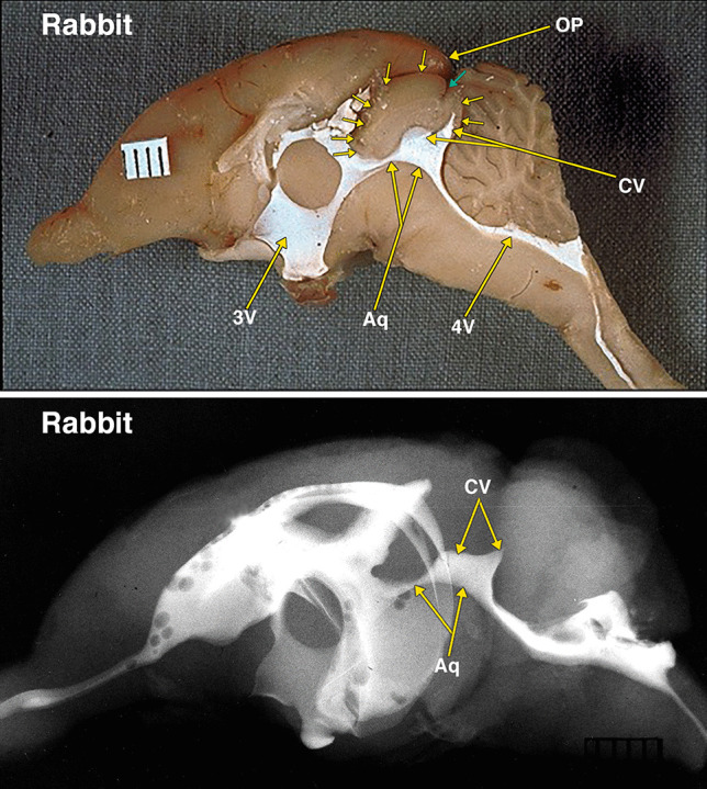
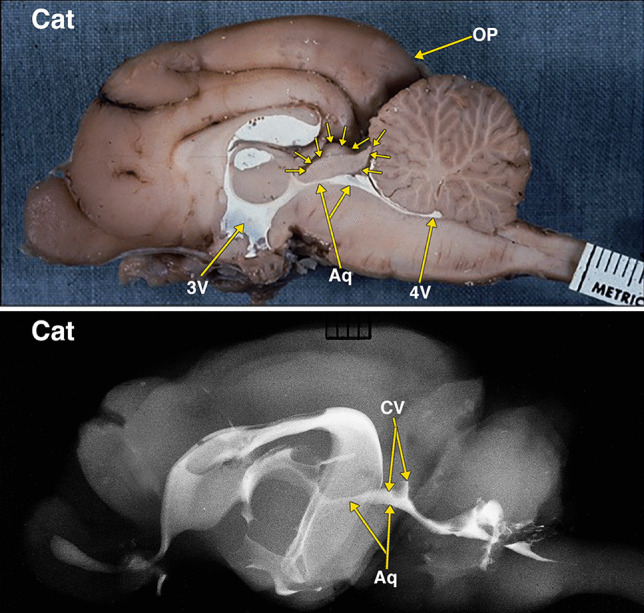
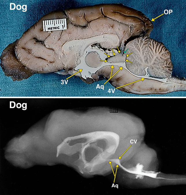
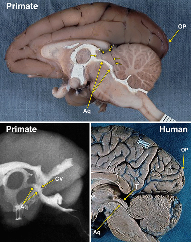
Photographs of brain dissections and radiographs of the cerebral ventricles and arteries of adult shark, frog, iguana, rabbit, cat, dog, and primate specimens, containing a barium-gelatin radiopaque compound, were analyzed focusing on the aqueduct, the optic ventricles, the quadrigeminal plate, and collicular ventricles. The anatomic information provided by the dissections and radiographs is not reproducible by any other radiopaque contrast currently available.
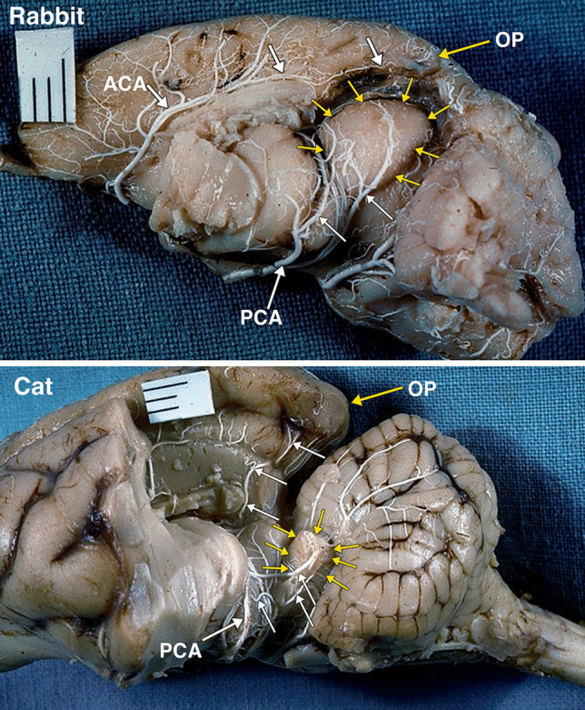
Dissected and radiographed cerebral ventricular and arterial systems of the vertebrates demonstrated midbrain tectum changes, including relative size modifications of the mammalian components of the tectum, simultaneously with the enlargement of the occipital lobe. There is a transformation of pre-mammalian optic ventricles to what appear to be collicular ventricles in mammals, as the aqueduct and collicular ventricle form a continuous cavity.
The mammalian tectum undergoes an evolutionary cephalization process consisting of relative size changes of the midbrain tectum structures. This is associated with enlargement of the occipital lobe, as part of overall neocortical expansion. Potentially, aqueductal anomalies could be explained by evolutionary modifications.
大量文献提出了导水管狭窄的多种病因。没有发现讨论进化改变可能解释导水管异常的出版物。本研究的目的是回顾脊椎动物脑桥结构的进化改变,这些改变可能解释人类导水管异常。由于获取脊椎动物材料的限制,目前进行脊椎动物比较研究是不可行的。因此,分析了 20 世纪 70 年代早期收集、注射、解剖和拍摄的脊椎动物材料,重点是导水管和中脑脑桥的成分。
分析了含有钡胶不透射线化合物的成年鲨鱼、青蛙、鬣蜥、兔子、猫、狗和灵长类动物标本的脑解剖照片和脑室及动脉的 X 光照片,重点是导水管、视神经室、四叠体板和丘脑室。目前任何其他不透射线对比剂都无法重现解剖和 X 光照片提供的解剖信息。
脊椎动物的脑室和动脉系统的解剖和 X 光显示,中脑脑桥发生了变化,包括哺乳动物脑桥成分的相对大小改变,同时枕叶增大。前哺乳动物视神经室转化为哺乳动物的丘脑室,因为导水管和丘脑室形成了一个连续的腔。
哺乳动物的脑桥经历了一个进化的头化过程,包括中脑脑桥结构的相对大小变化。这与枕叶的增大有关,是整个新皮质扩张的一部分。导水管异常可能是进化改变的结果。