• 文献检索
  • 文档翻译
  • 深度研究
  • 学术资讯
  • Suppr Zotero 插件Zotero 插件
  • 邀请有礼
  • 套餐&价格
  • 历史记录
应用&插件
Suppr Zotero 插件Zotero 插件浏览器插件Mac 客户端Windows 客户端微信小程序
定价
高级版会员购买积分包购买API积分包
服务
文献检索文档翻译深度研究API 文档MCP 服务
关于我们
关于 Suppr公司介绍联系我们用户协议隐私条款
关注我们

Suppr 超能文献

核心技术专利:CN118964589B侵权必究
粤ICP备2023148730 号-1Suppr @ 2026

文献检索

告别复杂PubMed语法,用中文像聊天一样搜索,搜遍4000万医学文献。AI智能推荐,让科研检索更轻松。

立即免费搜索

文件翻译

保留排版,准确专业,支持PDF/Word/PPT等文件格式,支持 12+语言互译。

免费翻译文档

深度研究

AI帮你快速写综述,25分钟生成高质量综述,智能提取关键信息,辅助科研写作。

立即免费体验

孕期诊断的细胞学/组织学宫颈高级别发育异常产后消退的相关预测因素

Predictive Factors Involved in Postpartum Regressions of Cytological/Histological Cervical High-Grade Dysplasia Diagnosed during Pregnancy.

作者信息

Gomez Yvan, Balaya Vincent, Lepigeon Karine, Mathevet Patrice, Jacot-Guillarmod Martine

机构信息

Colposcopy Unit, Women-Mother-Child Department, Lausanne University Hospital, 1011 Lausanne, Switzerland.

Faculty of Biology and Medicine, University of Lausanne, 1015 Lausanne, Switzerland.

出版信息

J Clin Med. 2021 Nov 15;10(22):5319. doi: 10.3390/jcm10225319.

DOI:10.3390/jcm10225319
PMID:34830596
原文链接:https://pmc.ncbi.nlm.nih.gov/articles/PMC8618594/
Abstract

OBJECTIVE

The aim of this study was to describe the evolution of high-grade cervical dysplasia during pregnancy and the postpartum period and to determine factors associated with dysplasia regression.

METHODS

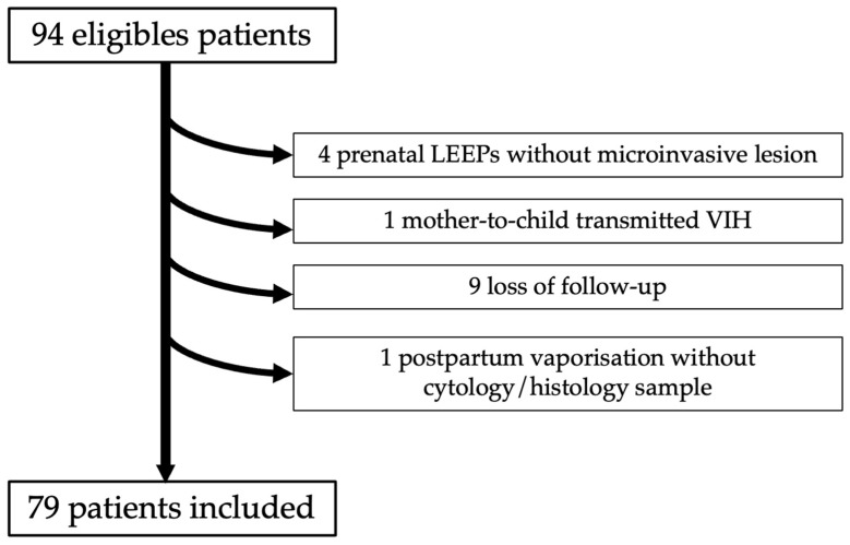
Pregnant patients diagnosed with high-grade lesions were identified in our tertiary hospital center. High-grade lesions were defined either cytologically, by high squamous intraepithelial lesion/atypical squamous cells being unable to exclude HSIL (HSIL/ASC-H), or histologically, with cervical intraepithelial neoplasia (CIN) 2+ (all CIN 2 and CIN 3) during pregnancy. Postpartum regression was defined cytologically or histologically by at least a one-degree reduction in severity from the antepartum diagnosis. A logistic regression model was applied to determine independent predictive factors for high-grade cervical dysplasia regression after delivery.

RESULTS

Between January 2000 and October 2017, 79 patients fulfilled the inclusion criteria and were analyzed. High-grade cervical lesions were diagnosed by cytology in 87% of cases (69/79) and confirmed by histology in 45% of those (31/69). The overall regression rate in our cohort was 43% (34/79). Univariate analysis revealed that parity ( = 0.04), diabetes ( = 0.04) and third trimester cytology ( = 0.009) were associated with dysplasia regression. Nulliparity (OR = 4.35; 95%CI = (1.03-18.42); = 0.046) was identified by multivariate analysis as an independent predictive factor of high-grade dysplasia regression. The presence of HSIL on third-trimester cervical cytology (OR = 0.17; 95%CI = (0.04-0.72); = 0.016) was identified as an independent predictive factor of high-grade dysplasia persistence at postpartum.

CONCLUSION

Our regression rate was high, at 43%, for high-grade cervical lesions postpartum. Parity status may have an impact on dysplasia regression during pregnancy. A cervical cytology should be performed at the third trimester to identify patients at risk of CIN persistence after delivery. However, larger cohorts are required to confirm these results.

摘要

目的

本研究旨在描述妊娠期间及产后高级别宫颈发育异常的演变情况,并确定与发育异常消退相关的因素。

方法

在我们的三级医院中心识别出诊断为高级别病变的孕妇。高级别病变通过细胞学定义为高度鳞状上皮内病变/不能排除高级别鳞状上皮内病变的非典型鳞状细胞(HSIL/ASC-H),或在组织学上定义为妊娠期间宫颈上皮内瘤变(CIN)2+(所有CIN 2和CIN 3)。产后消退通过细胞学或组织学定义为与产前诊断相比严重程度至少降低一级。应用逻辑回归模型确定分娩后高级别宫颈发育异常消退的独立预测因素。

结果

2000年1月至2017年10月,79例患者符合纳入标准并进行了分析。87%的病例(69/79)通过细胞学诊断为高级别宫颈病变,其中45%(31/69)经组织学证实。我们队列中的总体消退率为43%(34/79)。单因素分析显示,产次(P = 0.04)、糖尿病(P = 0.04)和孕晚期细胞学检查(P = 0.009)与发育异常消退相关。多因素分析确定未生育(OR = 4.35;95%CI =(1.03 - 18.42);P = 0.046)是高级别发育异常消退的独立预测因素。孕晚期宫颈细胞学检查存在HSIL(OR = 0.17;95%CI =(0.04 - 0.72);P = 0.016)被确定为产后高级别发育异常持续存在的独立预测因素。

结论

我们的产后高级别宫颈病变消退率较高,为43%。产次状态可能对妊娠期间发育异常的消退有影响。应在孕晚期进行宫颈细胞学检查,以识别分娩后有CIN持续存在风险的患者。然而,需要更大的队列来证实这些结果。

https://cdn.ncbi.nlm.nih.gov/pmc/blobs/dfa6/8618594/ad21df52927d/jcm-10-05319-g001.jpg

相似文献

1
Predictive Factors Involved in Postpartum Regressions of Cytological/Histological Cervical High-Grade Dysplasia Diagnosed during Pregnancy.孕期诊断的细胞学/组织学宫颈高级别发育异常产后消退的相关预测因素
J Clin Med. 2021 Nov 15;10(22):5319. doi: 10.3390/jcm10225319.
2
Diagnosis of Cervical Precancers by Endocervical Curettage at Colposcopy of Women With Abnormal Cervical Cytology.通过宫颈管刮术对宫颈细胞学异常女性进行阴道镜检查时诊断宫颈癌前病变
Obstet Gynecol. 2017 Dec;130(6):1218-1225. doi: 10.1097/AOG.0000000000002330.
3
Is the HSIL subclassification cytologically real and clinically justified?高级别鳞状上皮内病变(HSIL)的细胞学分型在细胞学上是否真实且在临床上是否合理?
Coll Antropol. 2010 Jun;34(2):395-400.
4
Absence of high-grade cervical intraepithelial neoplasia in conization specimens from patients with colposcopic biopsy-confirmed high-grade cervical intraepithelial neoplasia: Retrospective study of 1695 cases.阴道镜活检确诊为高级别宫颈上皮内瘤变患者的锥切标本中无高级别宫颈上皮内瘤变:1695例回顾性研究。
Front Oncol. 2022 Sep 14;12:980884. doi: 10.3389/fonc.2022.980884. eCollection 2022.
5
Is colposcopy warranted in women with external anogenital warts?外阴阴道湿疣患者行阴道镜检查是否合理?
J Low Genit Tract Dis. 2003 Jan;7(1):22-8. doi: 10.1097/00128360-200301000-00006.
6
p16 immunocytochemistry on cell blocks as an adjunct to cervical cytology: Potential reflex testing on specially prepared cell blocks from residual liquid-based cytology specimens.细胞块的p16免疫细胞化学作为宫颈细胞学的辅助手段:对来自剩余液基细胞学标本的特殊制备细胞块进行潜在的反射检测。
Cytojournal. 2011 Jan 31;8:1. doi: 10.4103/1742-6413.76379.
7
Triaging Atypical Squamous Cells-Cannot Exclude High-Grade Squamous Intraepithelial Lesion With p16/Ki67 Dual Stain.对非典型鳞状细胞进行分流——无法通过p16/Ki67双重染色排除高级别鳞状上皮内病变。
J Low Genit Tract Dis. 2017 Apr;21(2):108-111. doi: 10.1097/LGT.0000000000000297.
8
Risks of CIN 2+, CIN 3+, and Cancer by Cytology and Human Papillomavirus Status: The Foundation of Risk-Based Cervical Screening Guidelines.根据细胞学和人乳头瘤病毒状态评估CIN 2+、CIN 3+及癌症风险:基于风险的宫颈癌筛查指南的基础
J Low Genit Tract Dis. 2017 Oct;21(4):261-267. doi: 10.1097/LGT.0000000000000343.
9
The Positive Predictive Value of High-Grade Squamous Intraepithelial Lesion on Cytology for the Histological Diagnosis of Cervical Intraepithelial Neoplasia 2 or Higher: A Systematic Review.细胞学检查中高级别鳞状上皮内病变对宫颈上皮内瘤变2级或更高级别组织学诊断的阳性预测值:一项系统评价
Acta Cytol. 2019;63(3):206-214. doi: 10.1159/000497110. Epub 2019 Apr 4.
10
Cytology and high risk HPV testing in cervical cancer screening program: Outcome of 3-year follow-up in an academic institute.宫颈癌筛查项目中的细胞学和高危型人乳头瘤病毒检测:一所学术机构的3年随访结果
Diagn Cytopathol. 2018 Jan;46(1):22-27. doi: 10.1002/dc.23843. Epub 2017 Oct 19.

引用本文的文献

1
Antenatal and postnatal cervical precancer screening to increase coverage: experience from Battor, Ghana.增加宫颈癌前病变筛查覆盖率的产前和产后筛查:来自加纳巴托尔的经验
Ecancermedicalscience. 2023 Oct 30;17:1616. doi: 10.3332/ecancer.2023.1616. eCollection 2023.
2
The Fate of Cervical Dysplastic Lesions during Pregnancy and the Impact of the Delivery Mode: A Review.妊娠期间宫颈发育异常病变的转归及分娩方式的影响:综述
Cureus. 2023 Jul 18;15(7):e42100. doi: 10.7759/cureus.42100. eCollection 2023 Jul.
3
Characterization and clinical management of abnormal cytology findings in pregnant women: a retrospective analysis.

本文引用的文献

1
2019 ASCCP Risk-Based Management Consensus Guidelines for Abnormal Cervical Cancer Screening Tests and Cancer Precursors.2019年美国阴道镜和子宫颈病理学会基于风险的子宫颈癌筛查异常检测及癌前病变管理共识指南。
J Low Genit Tract Dis. 2020 Apr;24(2):102-131. doi: 10.1097/LGT.0000000000000525.
2
Immunobiology of Cervix Ripening.宫颈成熟的免疫生物学。
Front Immunol. 2020 Jan 24;10:3156. doi: 10.3389/fimmu.2019.03156. eCollection 2019.
3
Clinical outcome of high-grade cervical intraepithelial neoplasia during pregnancy: A 10-year experience.
对孕妇异常细胞学检查结果的特征描述和临床处理:回顾性分析。
Arch Gynecol Obstet. 2022 Dec;306(6):2017-2026. doi: 10.1007/s00404-022-06699-7. Epub 2022 Aug 17.
妊娠期高级别宫颈上皮内瘤变的临床转归:10 年经验。
Eur J Obstet Gynecol Reprod Biol. 2019 May;236:173-176. doi: 10.1016/j.ejogrb.2019.03.023. Epub 2019 Mar 25.
4
Natural History of Squamous Intraepithelial Lesions in Pregnancy and Mode of Delivery.妊娠期鳞状上皮内病变的自然史及分娩方式
Anticancer Res. 2018 Apr;38(4):2439-2442. doi: 10.21873/anticanres.12496.
5
Cervical intraepithelial neoplasia in pregnancy: Interference of pregnancy status with p16 and Ki-67 protein expression.妊娠期宫颈上皮内瘤变:妊娠状态对p16和Ki-67蛋白表达的影响
Oncol Lett. 2017 Jan;13(1):301-306. doi: 10.3892/ol.2016.5441. Epub 2016 Nov 29.
6
Natural history of cervical intraepithelial neoplasia in pregnancy: postpartum histo-pathologic outcome and review of the literature.妊娠期宫颈上皮内瘤变的自然史:产后组织病理学结局及文献综述
BMC Pregnancy Childbirth. 2016 Apr 7;16:74. doi: 10.1186/s12884-016-0861-8.
7
Colposcopically directed cervical biopsy during pregnancy; minor surgical and obstetrical complications and high rates of persistence and regression.阴道镜引导下的妊娠期宫颈活检;轻微的手术和产科并发症,以及持续性和退行性病变的高发生率。
Acta Obstet Gynecol Scand. 2013 Jun;92(6):692-9. doi: 10.1111/aogs.12138. Epub 2013 Apr 17.
8
High-grade cervical neoplasia during pregnancy: diagnosis, management and postpartum findings.妊娠期高级别宫颈病变:诊断、处理及产后发现。
Acta Obstet Gynecol Scand. 2013 Mar;92(3):293-7. doi: 10.1111/j.1600-0412.2012.01521.x.
9
Chemokine gene silencing in decidual stromal cells limits T cell access to the maternal-fetal interface.趋化因子基因沉默抑制了树突状基质细胞中 T 细胞向母体-胎儿界面的迁移。
Science. 2012 Jun 8;336(6086):1317-21. doi: 10.1126/science.1220030.
10
Mode of delivery influences the regression of abnormal cervical cytology.分娩方式影响异常宫颈细胞学的消退。
Gynecol Obstet Invest. 2011;72(4):234-8. doi: 10.1159/000324500. Epub 2011 Oct 20.