• 文献检索
  • 文档翻译
  • 深度研究
  • 学术资讯
  • Suppr Zotero 插件Zotero 插件
  • 邀请有礼
  • 套餐&价格
  • 历史记录
应用&插件
Suppr Zotero 插件Zotero 插件浏览器插件Mac 客户端Windows 客户端微信小程序
定价
高级版会员购买积分包购买API积分包
服务
文献检索文档翻译深度研究API 文档MCP 服务
关于我们
关于 Suppr公司介绍联系我们用户协议隐私条款
关注我们

Suppr 超能文献

核心技术专利:CN118964589B侵权必究
粤ICP备2023148730 号-1Suppr @ 2026

文献检索

告别复杂PubMed语法,用中文像聊天一样搜索,搜遍4000万医学文献。AI智能推荐,让科研检索更轻松。

立即免费搜索

文件翻译

保留排版,准确专业,支持PDF/Word/PPT等文件格式,支持 12+语言互译。

免费翻译文档

深度研究

AI帮你快速写综述,25分钟生成高质量综述,智能提取关键信息,辅助科研写作。

立即免费体验

[F]FMCH PET/CT生物标志物及相似性分析以完善寡转移前列腺癌的定义

[F]FMCH PET/CT biomarkers and similarity analysis to refine the definition of oligometastatic prostate cancer.

作者信息

Sollini Martina, Bartoli Francesco, Cavinato Lara, Ieva Francesca, Ragni Alessandra, Marciano Andrea, Zanca Roberta, Galli Luca, Paiar Fabiola, Pasqualetti Francesco, Erba Paola Anna

机构信息

Department of Biomedical Sciences, Humanitas University, Via Rita Levi Montalcini 4, Pieve Emanuele, Milan, Italy.

IRCCS Humanitas Research Hospital, Rozzano, Milan, Italy.

出版信息

EJNMMI Res. 2021 Nov 27;11(1):119. doi: 10.1186/s13550-021-00858-8.

DOI:10.1186/s13550-021-00858-8
PMID:34837532
原文链接:https://pmc.ncbi.nlm.nih.gov/articles/PMC8627538/
Abstract

BACKGROUND

The role of image-derived biomarkers in recurrent oligometastatic Prostate Cancer (PCa) is unexplored. This paper aimed to evaluate [F]FMCH PET/CT radiomic analysis in patients with recurrent PCa after primary radical therapy. Specifically, we tested intra-patient lesions similarity in oligometastatic and plurimetastatic PCa, comparing the two most used definitions of oligometastatic disease.

METHODS

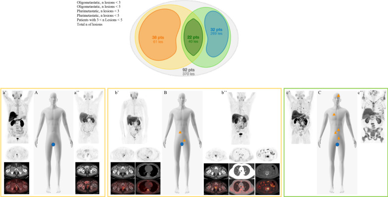
PCa patients eligible for [F]FMCH PET/CT presenting biochemical failure after first-line curative treatments were invited to participate in this prospective observational trial. PET/CT images of 92 patients were visually and quantitatively analyzed. Each patient was classified as oligometastatic or plurimetastatic according to the total number of detected lesions (up to 3 and up to 5 or > 3 and > 5, respectively). Univariate and intra-patient lesions' similarity analysis were performed.

RESULTS

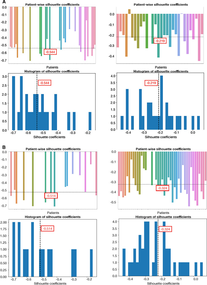
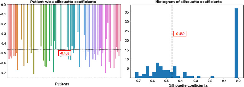
[F]FMCH PET/CT identified 370 lesions, anatomically classified as regional lymph nodes and distant metastases. Thirty-eight and 54 patients were designed oligometastatic and plurimetastatic, respectively, using a 3-lesion threshold. The number of oligometastic scaled up to 60 patients (thus 32 plurimetastatic patients) with a 5-lesion threshold. Similarity analysis showed high lesions' heterogeneity. Grouping patients according to the number of metastases, patients with oligometastatic PCa defined with a 5-lesion threshold presented lesions heterogeneity comparable to plurimetastic patients. Lesions within patients having a limited tumor burden as defined by three lesions were characterized by less heterogeneity.

CONCLUSIONS

We found a comparable heterogeneity between patients with up to five lesions and plurimetastic patients, while patients with up to three lesions were less heterogeneous than plurimetastatic patients, featuring different cells phenotypes in the two groups. Our results supported the use of a 3-lesion threshold to define oligometastatic PCa.

摘要

背景

影像衍生生物标志物在复发性寡转移前列腺癌(PCa)中的作用尚未得到探索。本文旨在评估[F]FMCH PET/CT放射组学分析在原发性根治性治疗后复发性PCa患者中的应用。具体而言,我们测试了寡转移和多转移PCa患者体内病变的相似性,比较了两种最常用的寡转移疾病定义。

方法

邀请一线根治性治疗后出现生化失败且符合[F]FMCH PET/CT检查条件的PCa患者参加这项前瞻性观察性试验。对92例患者的PET/CT图像进行了视觉和定量分析。根据检测到的病变总数(分别最多为3个和最多为5个或>3个和>5个)将每位患者分类为寡转移或多转移。进行了单变量和患者体内病变相似性分析。

结果

[F]FMCH PET/CT识别出370个病变,解剖学上分类为区域淋巴结和远处转移。使用3个病变阈值时,分别有38例和54例患者被定义为寡转移和多转移。使用5个病变阈值时,寡转移患者数量增至60例(因此多转移患者为32例)。相似性分析显示病变具有高度异质性。根据转移灶数量对患者进行分组,使用5个病变阈值定义的寡转移PCa患者的病变异质性与多转移患者相当。由3个病变定义的肿瘤负荷有限的患者体内的病变异质性较小。

结论

我们发现,病变数最多为5个的患者与多转移患者之间存在相当的异质性,而病变数最多为3个的患者比多转移患者的异质性小,两组具有不同的细胞表型。我们的结果支持使用3个病变阈值来定义寡转移PCa。

https://cdn.ncbi.nlm.nih.gov/pmc/blobs/ece7/8627538/52377c2d16ab/13550_2021_858_Fig3_HTML.jpg
https://cdn.ncbi.nlm.nih.gov/pmc/blobs/ece7/8627538/6591e070f491/13550_2021_858_Fig1_HTML.jpg
https://cdn.ncbi.nlm.nih.gov/pmc/blobs/ece7/8627538/ca460ec63763/13550_2021_858_Fig2_HTML.jpg
https://cdn.ncbi.nlm.nih.gov/pmc/blobs/ece7/8627538/52377c2d16ab/13550_2021_858_Fig3_HTML.jpg
https://cdn.ncbi.nlm.nih.gov/pmc/blobs/ece7/8627538/6591e070f491/13550_2021_858_Fig1_HTML.jpg
https://cdn.ncbi.nlm.nih.gov/pmc/blobs/ece7/8627538/ca460ec63763/13550_2021_858_Fig2_HTML.jpg
https://cdn.ncbi.nlm.nih.gov/pmc/blobs/ece7/8627538/52377c2d16ab/13550_2021_858_Fig3_HTML.jpg

相似文献

1
[F]FMCH PET/CT biomarkers and similarity analysis to refine the definition of oligometastatic prostate cancer.[F]FMCH PET/CT生物标志物及相似性分析以完善寡转移前列腺癌的定义
EJNMMI Res. 2021 Nov 27;11(1):119. doi: 10.1186/s13550-021-00858-8.
2
[F]Fluorocholine PET/CT-guided stereotactic body radiotherapy in patients with recurrent oligometastatic prostate cancer.[F]氟代胆碱PET/CT引导下的立体定向体部放射治疗复发性寡转移前列腺癌患者。
Eur J Nucl Med Mol Imaging. 2020 Jan;47(1):185-191. doi: 10.1007/s00259-019-04482-6. Epub 2019 Oct 16.
3
More advantages in detecting bone and soft tissue metastases from prostate cancer using F-PSMA PET/CT.使用F-PSMA PET/CT检测前列腺癌骨和软组织转移方面有更多优势。
Hell J Nucl Med. 2019 Jan-Apr;22(1):6-9. doi: 10.1967/s002449910952. Epub 2019 Mar 7.
4
Radiomics-Based Inter-Lesion Relation Network to Describe [F]FMCH PET/CT Imaging Phenotypes in Prostate Cancer.基于影像组学的病灶间关系网络用于描述前列腺癌中[F]FMCH PET/CT成像表型
Cancers (Basel). 2023 Jan 29;15(3):823. doi: 10.3390/cancers15030823.
5
[(18)F]Choline PET/CT and stereotactic body radiotherapy on treatment decision making of oligometastatic prostate cancer patients: preliminary results.[18F]胆碱正电子发射断层显像/计算机断层扫描与立体定向体部放疗在寡转移前列腺癌患者治疗决策中的应用:初步结果
Radiat Oncol. 2016 Jan 22;11:9. doi: 10.1186/s13014-016-0586-x.
6
Pelvic lymph node dissection for nodal oligometastatic prostate cancer detected by 68Ga-PSMA-positron emission tomography/computerized tomography.通过68Ga-PSMA正电子发射断层扫描/计算机断层扫描检测到的淋巴结寡转移前列腺癌的盆腔淋巴结清扫术
Prostate. 2015 Dec;75(16):1934-40. doi: 10.1002/pros.23091. Epub 2015 Sep 10.
7
Prostate-specific membrane antigen-positron emission tomography/computed tomography (PSMA-PET/CT)-guided stereotactic ablative body radiotherapy for oligometastatic prostate cancer: a single-institution experience and review of the published literature.前列腺特异性膜抗原正电子发射断层扫描/计算机断层扫描(PSMA-PET/CT)引导下的立体定向消融体部放射治疗寡转移前列腺癌:单机构经验及已发表文献综述
BJU Int. 2019 Nov;124 Suppl 1:19-30. doi: 10.1111/bju.14886. Epub 2019 Sep 11.
8
F-fluorodeoxyglucose positron emission tomography/computed tomography (PET/CT) is accurate for high-grade prostate cancer bone staging when compared to bone scintigraphy.与骨闪烁显像相比,F-氟脱氧葡萄糖正电子发射断层扫描/计算机断层扫描(PET/CT)对高级别前列腺癌骨分期的诊断准确。
Can Urol Assoc J. 2021 Oct;15(10):301-307. doi: 10.5489/cuaj.7107.
9
Feasibility of 18F-fluoromethylcholine PET/CT for imaging of vessel wall alterations in humans--first results.18F-氟甲基胆碱PET/CT用于人体血管壁改变成像的可行性——初步结果
Eur J Nucl Med Mol Imaging. 2008 Apr;35(4):815-20. doi: 10.1007/s00259-007-0685-x. Epub 2008 Jan 6.
10
ML Models Built Using Clinical Parameters and Radiomic Features Extracted from F-Choline PET/CT for the Prediction of Biochemical Recurrence after Metastasis-Directed Therapy in Patients with Oligometastatic Prostate Cancer.利用从F-胆碱PET/CT中提取的临床参数和影像组学特征构建的机器学习模型,用于预测寡转移前列腺癌患者转移导向治疗后的生化复发。
Diagnostics (Basel). 2024 Jun 15;14(12):1264. doi: 10.3390/diagnostics14121264.

引用本文的文献

1
Can Machine Learning Models Detect and Predict Lymph Node Involvement in Prostate Cancer? A Comprehensive Systematic Review.机器学习模型能否检测和预测前列腺癌的淋巴结转移?一项全面的系统评价。
J Clin Med. 2023 Nov 10;12(22):7032. doi: 10.3390/jcm12227032.
2
Radiomics-Based Inter-Lesion Relation Network to Describe [F]FMCH PET/CT Imaging Phenotypes in Prostate Cancer.基于影像组学的病灶间关系网络用于描述前列腺癌中[F]FMCH PET/CT成像表型
Cancers (Basel). 2023 Jan 29;15(3):823. doi: 10.3390/cancers15030823.
3
Oligometastatic Prostate Cancer: Current Status and Future Challenges.

本文引用的文献

1
Artificial intelligence and hybrid imaging: the best match for personalized medicine in oncology.人工智能与混合成像:肿瘤个性化医疗的最佳匹配
Eur J Hybrid Imaging. 2020 Dec 9;4(1):24. doi: 10.1186/s41824-020-00094-8.
2
Methodological framework for radiomics applications in Hodgkin's lymphoma.霍奇金淋巴瘤中放射组学应用的方法学框架。
Eur J Hybrid Imaging. 2020 Jun 1;4(1):9. doi: 10.1186/s41824-020-00078-8.
3
Metabolic regulation of prostate cancer heterogeneity and plasticity.前列腺癌异质性和可塑性的代谢调控
寡转移前列腺癌:现状与未来挑战。
J Nucl Med. 2022 Nov;63(11):1628-1635. doi: 10.2967/jnumed.121.263124.
4
The Road to Dissemination: The Concept of Oligometastases and the Barriers for Widespread Disease.传播之路:寡转移的概念及广泛疾病的障碍
Cancers (Basel). 2022 Apr 18;14(8):2046. doi: 10.3390/cancers14082046.
Semin Cancer Biol. 2022 Jul;82:94-119. doi: 10.1016/j.semcancer.2020.12.002. Epub 2020 Dec 5.
4
Climbing the steps of the evidence-based medicine pyramid: highlights from Annals of Nuclear Medicine 2019.攀登循证医学金字塔的台阶:2019 年《核医学年鉴》的亮点。
Eur J Nucl Med Mol Imaging. 2021 May;48(5):1293-1301. doi: 10.1007/s00259-020-05073-6. Epub 2020 Nov 4.
5
EAU-EANM-ESTRO-ESUR-SIOG Guidelines on Prostate Cancer. Part II-2020 Update: Treatment of Relapsing and Metastatic Prostate Cancer.EAU-EANM-ESTRO-ESUR-SIOG 前列腺癌诊治指南。第二部分-2020 年更新:复发性和转移性前列腺癌的治疗。
Eur Urol. 2021 Feb;79(2):263-282. doi: 10.1016/j.eururo.2020.09.046. Epub 2020 Oct 7.
6
Imaging-Based Prediction of Molecular Therapy Targets in NSCLC by Radiogenomics and AI Approaches: A Systematic Review.基于影像组学和人工智能方法的非小细胞肺癌分子治疗靶点的影像预测:一项系统综述
Diagnostics (Basel). 2020 May 30;10(6):359. doi: 10.3390/diagnostics10060359.
7
The Image Biomarker Standardization Initiative: Standardized Quantitative Radiomics for High-Throughput Image-based Phenotyping.影像生物标志物标准化倡议:高通量基于影像表型的标准化定量放射组学。
Radiology. 2020 May;295(2):328-338. doi: 10.1148/radiol.2020191145. Epub 2020 Mar 10.
8
Spatial modeling of prostate cancer metabolic gene expression reveals extensive heterogeneity and selective vulnerabilities.前列腺癌代谢基因表达的空间建模揭示了广泛的异质性和选择性脆弱性。
Sci Rep. 2020 Feb 26;10(1):3490. doi: 10.1038/s41598-020-60384-w.
9
Imaging for Metastasis in Prostate Cancer: A Review of the Literature.前列腺癌转移的影像学检查:文献综述
Front Oncol. 2020 Jan 31;10:55. doi: 10.3389/fonc.2020.00055. eCollection 2020.
10
Quantitative imaging biomarkers in nuclear medicine: from SUV to image mining studies. Highlights from annals of nuclear medicine 2018.核医学中的定量影像生物标志物:从 SUV 到影像挖掘研究。2018 年核医学纪事亮点。
Eur J Nucl Med Mol Imaging. 2019 Dec;46(13):2737-2745. doi: 10.1007/s00259-019-04531-0. Epub 2019 Nov 5.