Department of Neurological Surgery, University of Texas Southwestern Medical Center, Dallas, Texas, USA.
O'Donnell Brain Institute, University of Texas Southwestern Medical Center, Dallas, Texas,USA.
Neuro Oncol. 2022 Apr 1;24(4):612-623. doi: 10.1093/neuonc/noab273.
Historically, creating patient-derived models of lower-grade glioma (LGG) has been challenging, contributing to few experimental platforms that support laboratory-based investigations of this disease. Although organoid modeling approaches have recently been employed to create in vitro models of high-grade glioma (HGG), it is unknown whether this approach can be successfully applied to LGG.
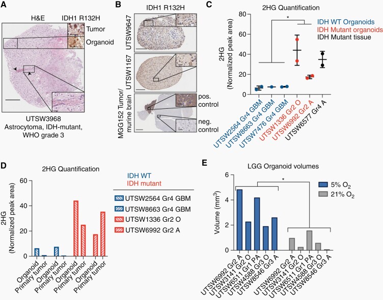
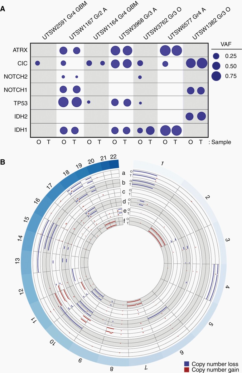
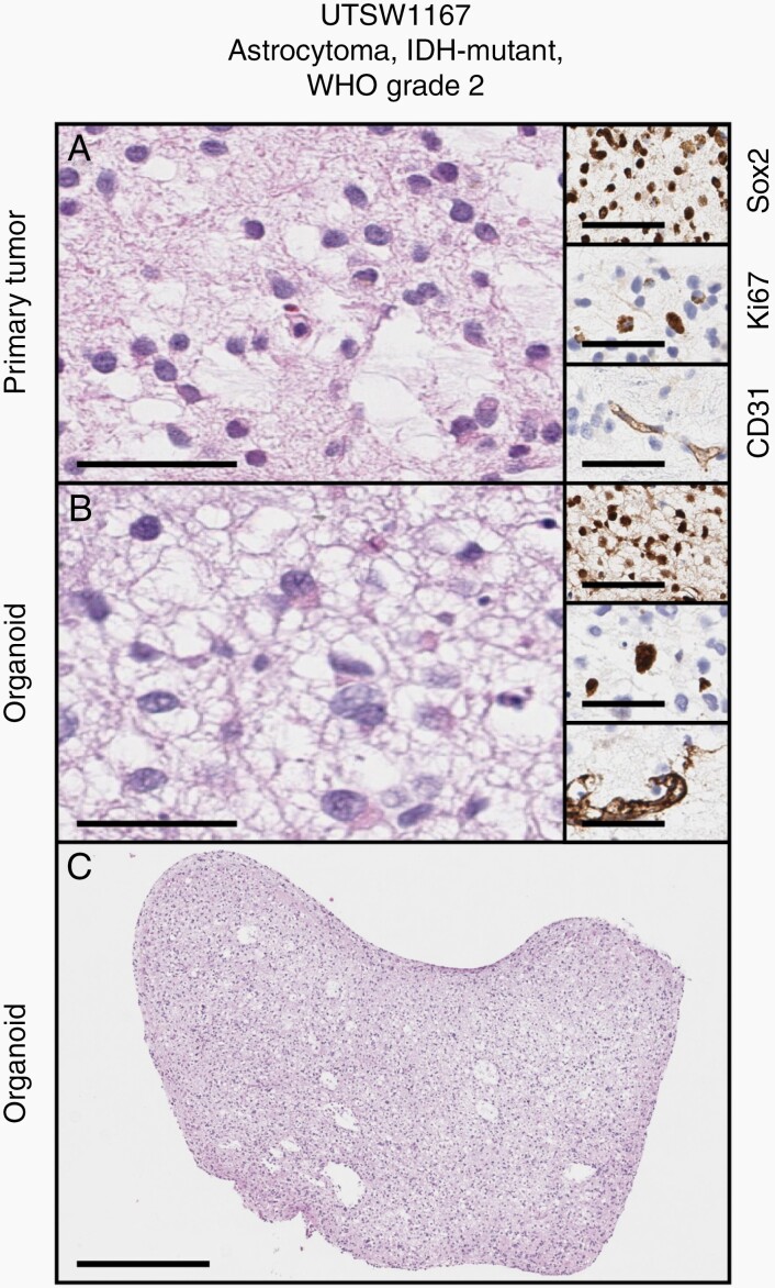
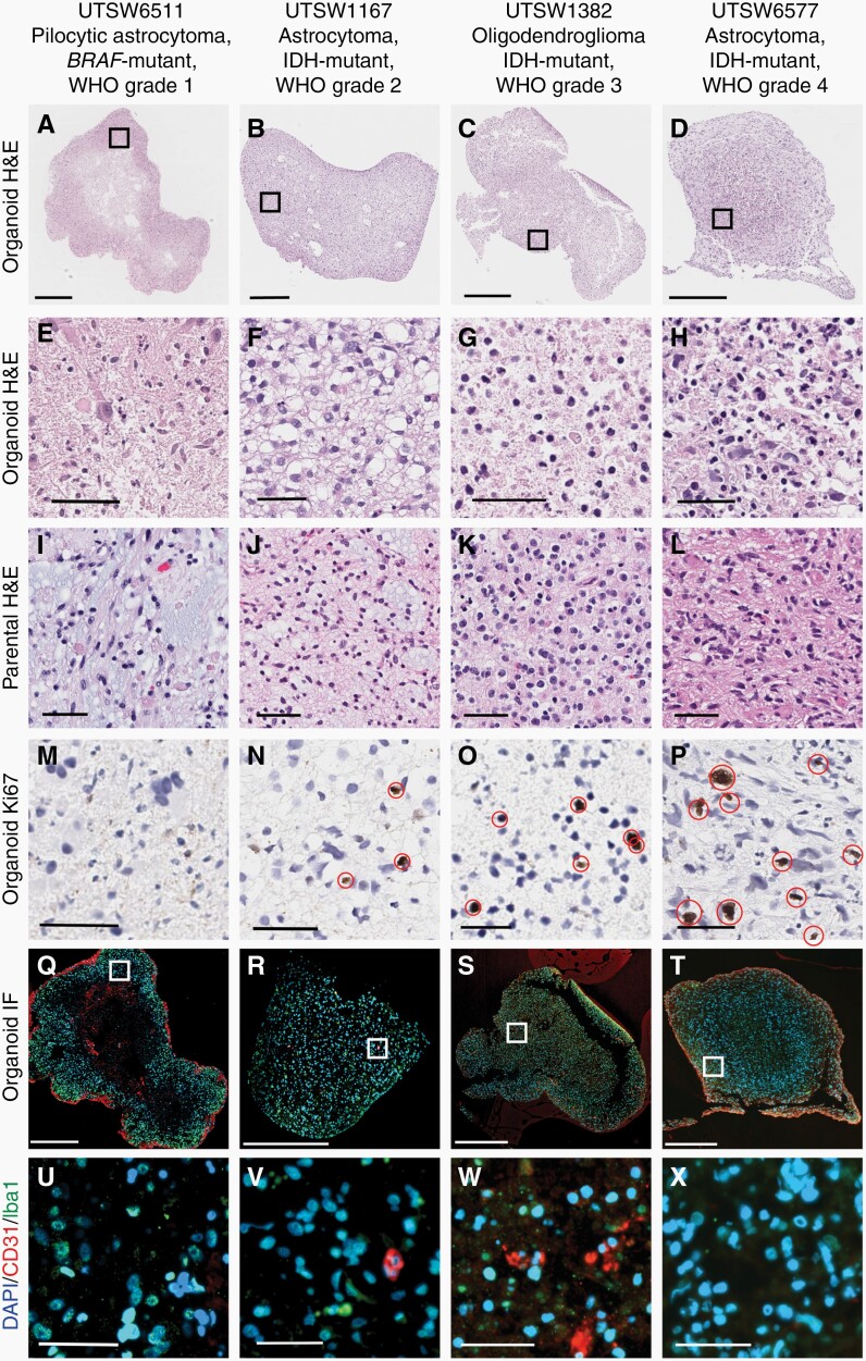
In this study, we developed an optimized protocol for the establishment of organoids from LGG primary tissue samples by utilizing physiologic (5%) oxygenation conditions and employed it to produce the first known suite of these models. To assess their fidelity, we surveyed key biological features of patient-derived organoids using metabolic, genomic, histologic, and lineage marker gene expression assays.
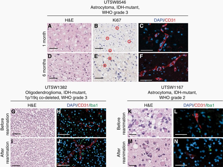
Organoid models were created with a success rate of 91% (n = 20/22) from primary tumor samples across glioma histological subtypes and tumor grades (WHO Grades 1-4), and a success rate of 87% (13/15) for WHO Grade 1-3 tumors. Patient-derived organoids recapitulated stemness, proliferative, and tumor-stromal composition profiles of their respective parental tumor specimens. Cytoarchitectural, mutational, and metabolic traits of parental tumors were also conserved. Importantly, LGG organoids were maintained in vitro for weeks to months and reanimated after biobanking without loss of integrity.
We report an efficient method for producing faithful in vitro models of LGG. New experimental platforms generated through this approach are well positioned to support preclinical studies of this disease, particularly those related to tumor immunology, tumor-stroma interactions, identification of novel drug targets, and personalized assessments of treatment response profiles.
从历史上看,构建低级别胶质瘤(LGG)的患者衍生模型一直具有挑战性,这导致很少有实验平台能够支持对这种疾病的实验室研究。尽管类器官建模方法最近已被用于创建高级别胶质瘤(HGG)的体外模型,但尚不清楚该方法是否可以成功应用于 LGG。
在这项研究中,我们通过利用生理(5%)氧合条件开发了一种从 LGG 原发组织样本中建立类器官的优化方案,并利用该方案首次成功建立了此类模型。为了评估它们的保真度,我们使用代谢、基因组、组织学和谱系标记基因表达分析来调查患者衍生类器官的关键生物学特征。
从各种胶质瘤组织亚型和肿瘤分级(WHO 分级 1-4)的原发肿瘤样本中,类器官模型的成功率为 91%(n=20/22),而 WHO 分级 1-3 肿瘤的成功率为 87%(n=13/15)。患者衍生的类器官再现了其各自母肿瘤标本的干性、增殖和肿瘤-基质组成特征。亲本肿瘤的细胞结构、突变和代谢特征也得到了保留。重要的是,LGG 类器官可以在体外维持数周到数月,并在生物银行储存后重新激活而不会失去完整性。
我们报告了一种高效的 LGG 体外模型构建方法。通过这种方法生成的新实验平台非常适合支持该疾病的临床前研究,特别是那些与肿瘤免疫学、肿瘤-基质相互作用、新药物靶点的鉴定以及治疗反应谱的个性化评估相关的研究。