Department of Radiology, Samsung Medical Center, Sungkyunkwan University School of Medicine, 81 Irwon-ro, Gangnam-gu, 06351, Seoul, Korea.
Philips Korea, Seoul, Korea.
Sci Rep. 2022 Jan 7;12(1):73. doi: 10.1038/s41598-021-03928-y.
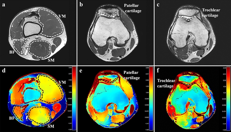
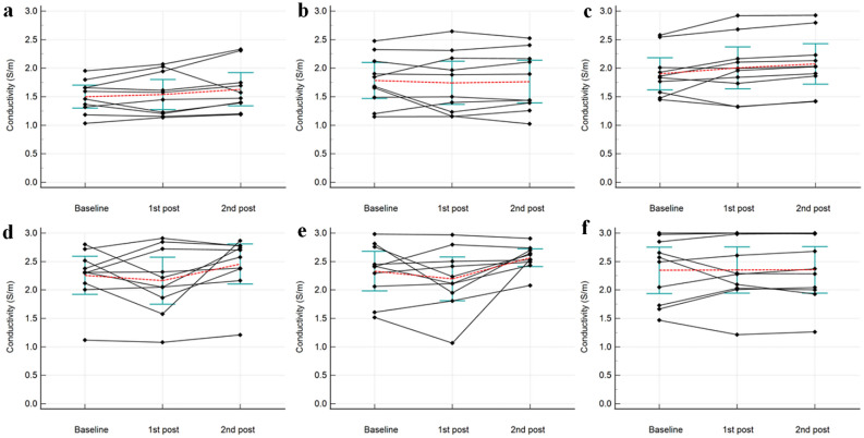
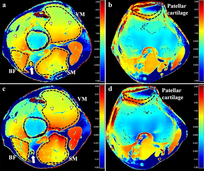
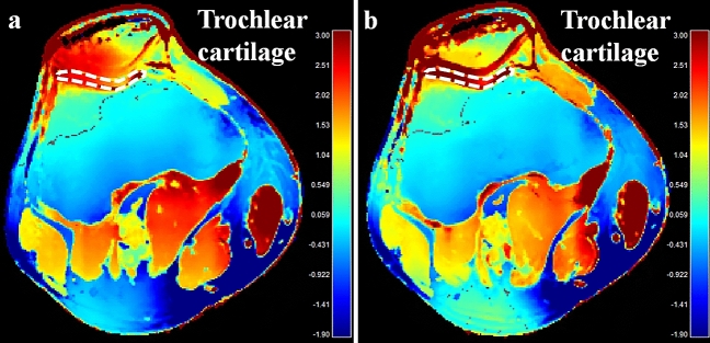
This study aimed to investigate whether in vivo MR-electrical properties tomography (MR-EPT) is feasible in musculoskeletal tissues by evaluating the conductivity of muscle, cartilage, and peripheral nerve around the knee joint, and to explore whether these measurements change after exercise. This prospective study was approved by the institutional review board. On February 2020, ten healthy volunteers provided written informed consent and underwent MRI of the right knee using a three-dimensional balanced steady-state free precession (bSSFP) sequence. To test the effect of loading, the subjects performed 60 squatting exercises after baseline MRI, immediately followed by post-exercise MRI with the same sequences. After reconstruction of conductivity map based on the bSSFP sequence, conductivity of muscles, cartilages, and nerves were measured. Measurements between the baseline and post-exercise MRI were compared using the paired t-test. Test-retest reliability for baseline conductivity was evaluated using the intraclass correlation coefficient. The baseline and post-exercise conductivity values (mean ± standard deviation) [S/m] of muscles, cartilages, and nerves were 1.73 ± 0.40 and 1.82 ± 0.50 (p = 0.048), 2.29 ± 0.47 and 2.51 ± 0.37 (p = 0.006), and 2.35 ± 0.57 and 2.36 ± 0.57 (p = 0.927), respectively. Intraclass correlation coefficient for the baseline conductivity of muscles, cartilages, and nerves were 0.89, 0.67, and 0.89, respectively. In conclusion, in vivo conductivity measurement of musculoskeletal tissues is feasible using MR-EPT. Conductivity of muscles and cartilages significantly changed with an overall increase after exercise.
本研究旨在通过评估膝关节周围肌肉、软骨和周围神经的电导率,来探究在肌肉骨骼组织中进行体内磁共振电特性断层成像(MR-EPT)是否可行,并探讨这些测量值在运动后是否发生变化。本前瞻性研究得到了机构审查委员会的批准。2020 年 2 月,10 名健康志愿者书面同意并接受了右侧膝关节的磁共振成像检查,使用三维平衡稳态自由进动(bSSFP)序列。为了测试加载的效果,受试者在基线 MRI 后进行了 60 次深蹲运动,随后立即进行了相同序列的运动后 MRI。基于 bSSFP 序列重建电导率图后,测量肌肉、软骨和神经的电导率。使用配对 t 检验比较基线和运动后 MRI 的测量值。使用组内相关系数评估基线电导率的测试-重测信度。肌肉、软骨和神经的基线和运动后电导率值(平均值±标准差)[S/m]分别为 1.73±0.40 和 1.82±0.50(p=0.048)、2.29±0.47 和 2.51±0.37(p=0.006)以及 2.35±0.57 和 2.36±0.57(p=0.927)。肌肉、软骨和神经的基线电导率的组内相关系数分别为 0.89、0.67 和 0.89。总之,使用 MR-EPT 对肌肉骨骼组织进行体内电导率测量是可行的。肌肉和软骨的电导率在运动后整体增加,且明显发生变化。