Korzowski Andreas, Weckesser Nina, Franke Vanessa L, Breitling Johannes, Goerke Steffen, Schlemmer Heinz-Peter, Ladd Mark E, Bachert Peter, Paech Daniel
Department of Medical Physics in Radiology, German Cancer Research Center (DKFZ), Heidelberg, Germany.
Department of Radiology, German Cancer Research Center (DKFZ), Heidelberg, Germany.
Front Neurol. 2021 Dec 23;12:735071. doi: 10.3389/fneur.2021.735071. eCollection 2021.
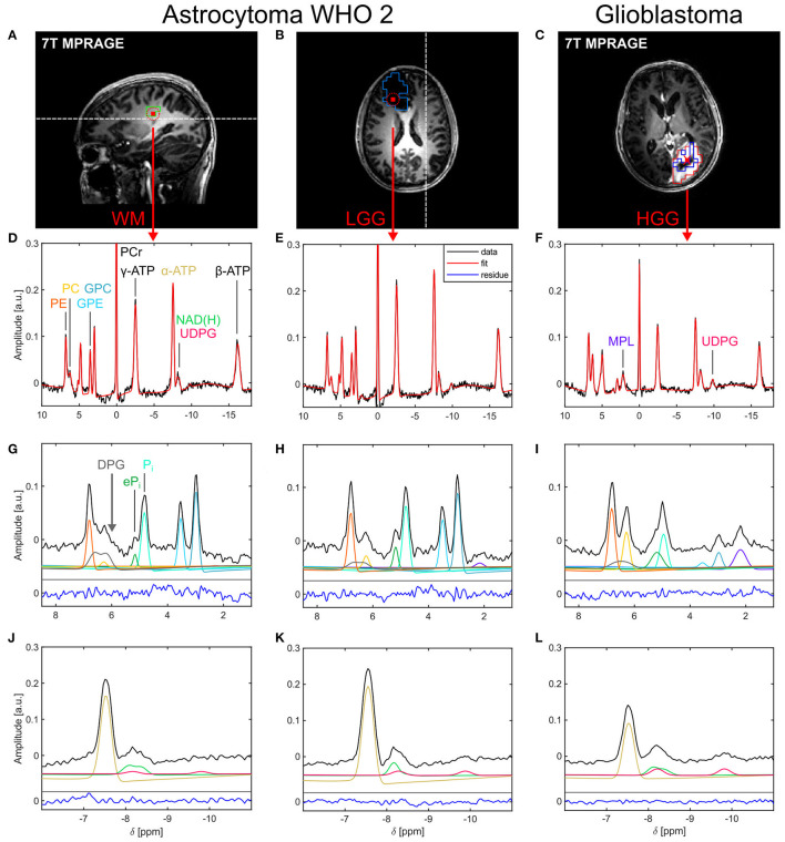
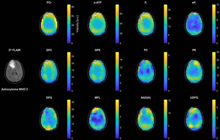
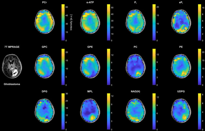
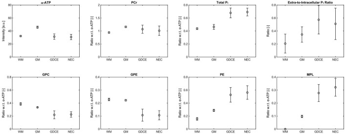
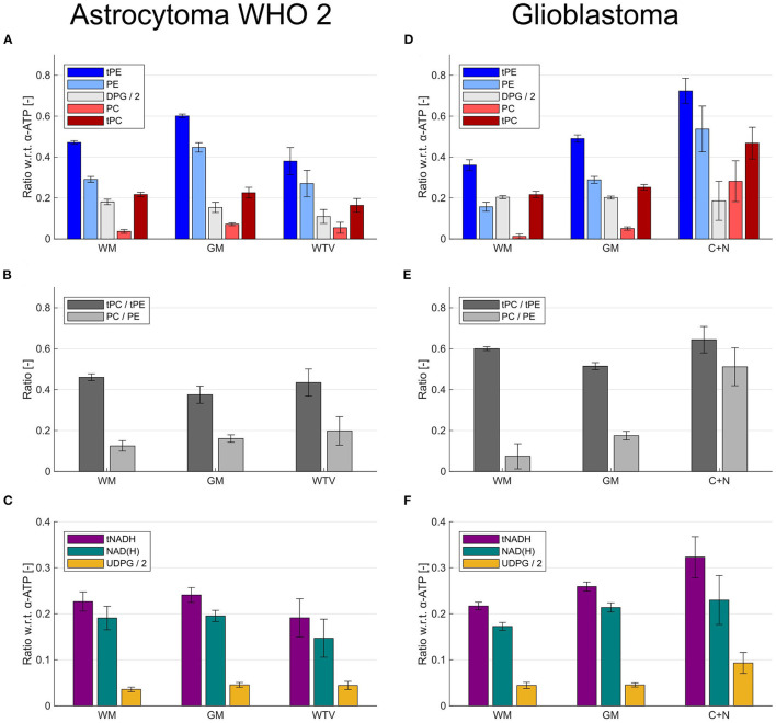
Phosphorus magnetic resonance spectroscopic imaging (P MRSI) is of particular interest for investigations of patients with brain tumors as it enables to non-invasively assess altered energy and phospholipid metabolism . However, the limited sensitivity of P MRSI hampers its broader application at clinical field strengths. This study aimed to identify the additional value of P MRSI in patients with glioma at ultra-high = 7T, where the increase in signal-to-noise ratio may foster its applicability for clinical research. High-quality, 3D P MRSI datasets with an effective voxel size of 5.7 ml were acquired from the brains of seven patients with newly diagnosed glioma. An optimized quantification model was implemented to reliably extract an extended metabolic profile, including low-concentrated metabolites such as extracellular inorganic phosphate, nicotinamide adenine dinucleotide [NAD(H)], and uridine diphosphoglucose (UDPG), which may act as novel tumor markers; a background signal was extracted as well, which affected measures of phosphomonoesters beneficially. Application of this model to the MRSI datasets yielded high-resolution maps of 12 different P metabolites, showing clear metabolic differences between white matter (WM) and gray matter, and between healthy and tumor tissues. Moreover, differences between tumor compartments in patients with high-grade glioma (HGG), i.e., gadolinium contrast-enhancing/necrotic regions (C+N) and peritumoral edema, could also be suggested from these maps. In the group of patients with HGG, the most significant changes in metabolite intensities were observed in C+N compared to WM, i.e., for phosphocholine +340%, UDPG +54%, glycerophosphoethanolamine -45%, and adenosine-5'-triphosphate -29%. Furthermore, a prominent signal from mobile phospholipids appeared in C+N. In the group of patients with low-grade glioma, only the NAD(H) intensity changed significantly by -28% in the tumor compared to WM. Besides the potential of P MRSI at 7T to provide novel insights into the biochemistry of gliomas , the attainable spatial resolutions improve the interpretability of P metabolite intensities obtained from malignant tissues, particularly when only subtle differences compared to healthy tissues are expected. In conclusion, this pilot study demonstrates that P MRSI at 7T has potential value for the clinical research of glioma.
磷磁共振波谱成像(P MRSI)对于脑肿瘤患者的研究尤为重要,因为它能够无创地评估能量和磷脂代谢的改变。然而,P MRSI有限的灵敏度阻碍了其在临床场强下的更广泛应用。本研究旨在确定P MRSI在超高场强(7T)下对胶质瘤患者的附加价值,在该场强下信噪比的增加可能会促进其在临床研究中的适用性。从7例新诊断的胶质瘤患者的大脑中获取了有效体素大小为5.7毫升的高质量3D P MRSI数据集。实施了优化的定量模型,以可靠地提取扩展的代谢谱,包括细胞外无机磷酸盐、烟酰胺腺嘌呤二核苷酸[NAD(H)]和尿苷二磷酸葡萄糖(UDPG)等低浓度代谢物,这些代谢物可能作为新型肿瘤标志物;还提取了背景信号,其对磷酸单酯的测量有有益影响。将该模型应用于MRSI数据集产生了12种不同磷代谢物的高分辨率图谱,显示了白质(WM)和灰质之间以及健康组织和肿瘤组织之间明显的代谢差异。此外,从这些图谱中还可以看出高级别胶质瘤(HGG)患者肿瘤区域之间的差异,即钆增强/坏死区域(C+N)和瘤周水肿之间的差异。在HGG患者组中,与WM相比,C+N中代谢物强度的最显著变化被观察到,即磷酸胆碱增加340%,UDPG增加54%,甘油磷酸乙醇胺减少45%,三磷酸腺苷减少29%。此外,在C+N中出现了来自可移动磷脂的显著信号。在低级别胶质瘤患者组中,与WM相比,肿瘤中只有NAD(H)强度显著变化了28%。除了7T场强下P MRSI为胶质瘤生物化学提供新见解的潜力外,可实现的空间分辨率提高了从恶性组织获得的磷代谢物强度的可解释性,特别是当预期与健康组织只有细微差异时。总之,这项初步研究表明7T场强下的P MRSI对胶质瘤的临床研究具有潜在价值。