Azzolini Claudio, Donati Simone, Micheloni Giovanni, Moretti Vittoria, Valli Roberto, Acquati Francesco, Costantino Lucy, Ferrara Fulvio, Borroni Davide, Premi Elias, Testa Francesco, Simonelli Francesca, Porta Giovanni
Department of Medicine and Surgery, University of Insubria, Varese-Como, Italy.
Ophthalmology Unit, ASST Sette-Laghi, Varese, Italy.
J Ophthalmol. 2021 Dec 31;2021:6265553. doi: 10.1155/2021/6265553. eCollection 2021.
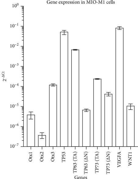
Müller glial cells typically activate to react to hypoxic tissue damage in several retinal diseases. We evaluated the response to a hypoxia-mimicking stimulus on the expression of a set of genes, known to contribute to eye morphogenesis and cell differentiation.
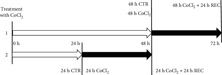
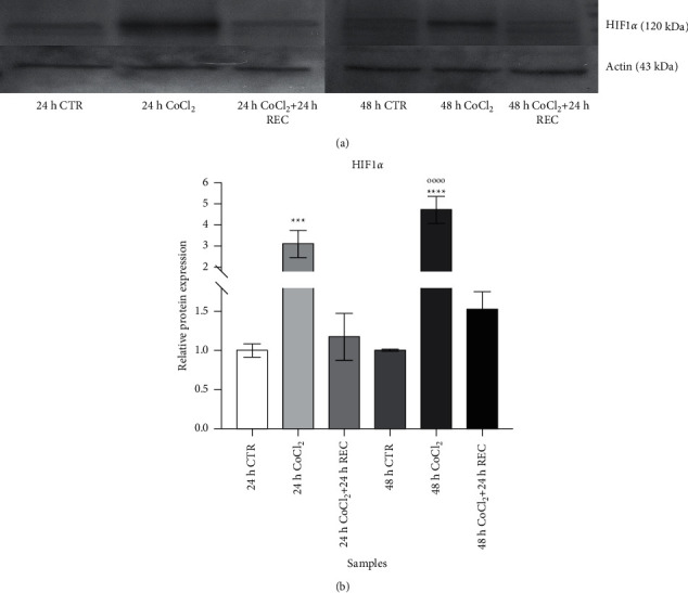
A MIO-M1 Müller cell line was cultured in a hypoxia-mimicking environment by the addition of cobalt chloride to the culture medium, followed by a recovery time in which we mimic restoration from the hypoxic insult. The HIF-1 protein and VEGF-A gene expression were quantified to verify the induction of a hypoxia-like state.
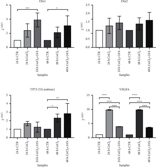
Among the genes under study, we did not observe any difference in the expression levels of and during treatment; conversely, was overexpressed during recovery steps. The VEGF-A gene was strongly upregulated at both the CoCl and recovery time points. The transactivated isoform (TA) of the gene showed an overexpression in long-term exposure to the hypoxic stimulus with a further increase after recovery. . Our molecular analysis is able to describe the activation of a set of genes, never before described, that can drive the response to a hypoxia-like status. The improved comprehension of these cellular events will be useful for designing new therapeutical approaches for retinal pathologies.
在几种视网膜疾病中,米勒胶质细胞通常会被激活以应对缺氧性组织损伤。我们评估了模拟缺氧刺激对一组已知有助于眼睛形态发生和细胞分化的基因表达的反应。
通过向培养基中添加氯化钴,在模拟缺氧的环境中培养MIO-M1米勒细胞系,随后进行恢复阶段,在此期间模拟从缺氧损伤中恢复的过程。对缺氧诱导因子-1(HIF-1)蛋白和血管内皮生长因子-A(VEGF-A)基因表达进行定量,以验证是否诱导出类似缺氧的状态。
在所研究的基因中,我们在处理过程中未观察到[具体基因1]和[具体基因2]表达水平有任何差异;相反,[具体基因3]在恢复阶段过表达。VEGF-A基因在氯化钴处理和恢复时间点均强烈上调。[具体基因4]的反式激活异构体(TA)在长期暴露于缺氧刺激时过表达,恢复后进一步增加。我们的分子分析能够描述一组此前从未描述过的基因的激活情况,这些基因可驱动对类似缺氧状态的反应。对这些细胞事件的深入理解将有助于设计针对视网膜病变的新治疗方法。