Andjelic Sofija, Drašlar Kazimir, Hvala Anastazija, Hawlina Marko
Eye Hospital, University Medical Centre, Ljubljana, Slovenia.
Department of Biology, Biotechnical Faculty, University of Ljubljana, Ljubljana, Slovenia.
Front Med (Lausanne). 2021 Dec 22;8:802275. doi: 10.3389/fmed.2021.802275. eCollection 2021.
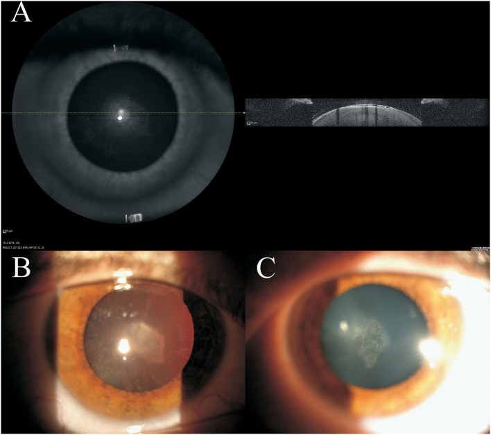
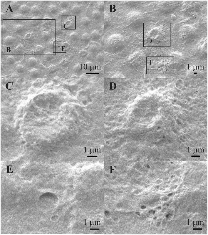
The purpose of this work is to examine the structure of the anterior lens epithelial cells (aLECs) of presenile idiopathic cortical cataract to investigate the possible structural reasons for its development. The anterior lens capsules (aLCs: basement membrane and associated lens epithelial cells) were obtained from routine uneventful cataract surgery of 5 presenile cataract patients (16 and 41 years old women and 29, 39, and 45 years old men). None of the patients had family history of cataract, medication, or trauma and they were otherwise healthy. In addition, the patients did not have any other abnormal features in the ocular status except cataract. The aLCs were prepared for scanning electron microscopy (SEM) and transmission electron microscopy (TEM). The most prominent abnormal features observed by SEM for all 5 studied presenile cataract patients were the changes of the aLECs structure with the dents, the selective concavity of some LECs, at their apical side centrally toward the nucleus. In addition, TEM showed the thinning of the lens epithelium with the segmentally concave cells and the compressed and elongated nuclei. Abnormal and distinguishable structural features were observed in the anterior lens epithelium aLECs in all 5 patients with presenile cataract. Disturbed structure of aLECs, regularly present in presenile cataract type is shown that might be associated with water accumulation in the presenile idiopathic cortical cataract lens.
这项工作的目的是检查早老性特发性皮质性白内障的前囊膜晶状体上皮细胞(aLECs)结构,以探究其发生发展的可能结构原因。从5例早老性白内障患者(2名16岁和41岁女性以及3名29岁、39岁和45岁男性)的常规白内障手术中获取前囊膜(aLCs:基底膜及相关晶状体上皮细胞)。所有患者均无白内障家族史、用药史或外伤史,且其他方面健康。此外,除白内障外,患者眼部状况无任何其他异常特征。对aLCs进行扫描电子显微镜(SEM)和透射电子显微镜(TEM)检查。在所有5例研究的早老性白内障患者中,SEM观察到的最显著异常特征是aLECs结构改变,出现凹痕,部分LECs顶端中央朝向细胞核处有选择性凹陷。此外,TEM显示晶状体上皮变薄,细胞呈节段性凹陷,细胞核受压且拉长。在所有5例早老性白内障患者的前囊膜晶状体上皮细胞(aLECs)中均观察到异常且可区分的结构特征。研究表明,早老性白内障类型中经常出现的aLECs结构紊乱可能与早老性特发性皮质性白内障晶状体中的水积聚有关。