• 文献检索
  • 文档翻译
  • 深度研究
  • 学术资讯
  • Suppr Zotero 插件Zotero 插件
  • 邀请有礼
  • 套餐&价格
  • 历史记录
应用&插件
Suppr Zotero 插件Zotero 插件浏览器插件Mac 客户端Windows 客户端微信小程序
定价
高级版会员购买积分包购买API积分包
服务
文献检索文档翻译深度研究API 文档MCP 服务
关于我们
关于 Suppr公司介绍联系我们用户协议隐私条款
关注我们

Suppr 超能文献

核心技术专利:CN118964589B侵权必究
粤ICP备2023148730 号-1Suppr @ 2026

文献检索

告别复杂PubMed语法,用中文像聊天一样搜索,搜遍4000万医学文献。AI智能推荐,让科研检索更轻松。

立即免费搜索

文件翻译

保留排版,准确专业,支持PDF/Word/PPT等文件格式,支持 12+语言互译。

免费翻译文档

深度研究

AI帮你快速写综述,25分钟生成高质量综述,智能提取关键信息,辅助科研写作。

立即免费体验

自噬相关的肝星状细胞激活通过促进其囊泡分泌来减少细胞 miR-29a。

Autophagy-Related Activation of Hepatic Stellate Cells Reduces Cellular miR-29a by Promoting Its Vesicular Secretion.

机构信息

Institute for Pathology, Medical Faculty and University Hospital of Cologne, University of Cologne, Cologne, Germany; Center for Molecular Medicine Cologne (CMMC), University of Cologne, Cologne, Germany.

Institute for Pathology, Medical Faculty and University Hospital of Cologne, University of Cologne, Cologne, Germany.

出版信息

Cell Mol Gastroenterol Hepatol. 2022;13(6):1701-1716. doi: 10.1016/j.jcmgh.2022.02.013. Epub 2022 Feb 24.

DOI:
10.1016/j.jcmgh.2022.02.013
PMID:35219894
原文链接:https://pmc.ncbi.nlm.nih.gov/articles/PMC9046234/
Abstract

BACKGROUND & AIMS: Liver fibrosis arises from long-term chronic liver injury, accompanied by an accelerated wound healing response with interstitial accumulation of extracellular matrix (ECM). Activated hepatic stellate cells (HSC) are the main source for ECM production. MicroRNA29a (miR-29a) is a crucial antifibrotic miRNA that is repressed during fibrosis, resulting in up-regulation of collagen synthesis.

METHODS

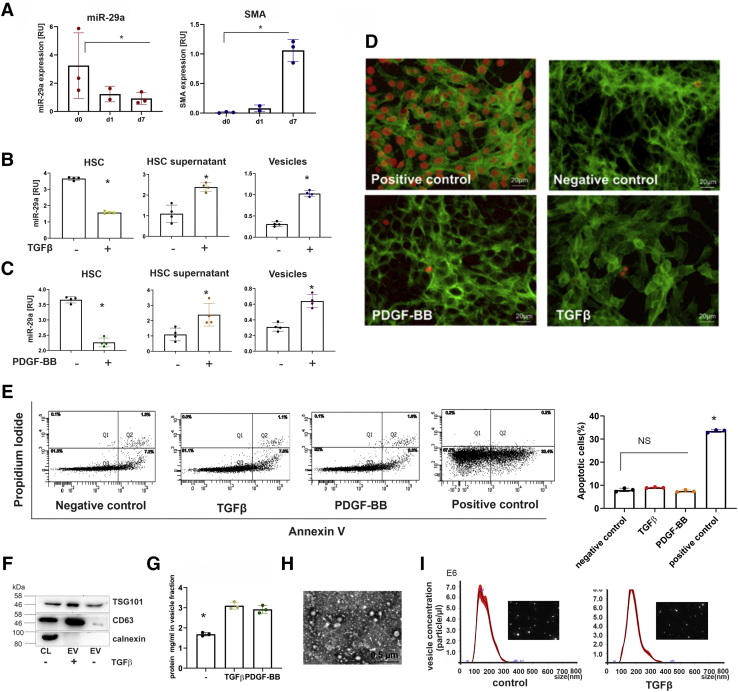
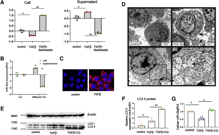
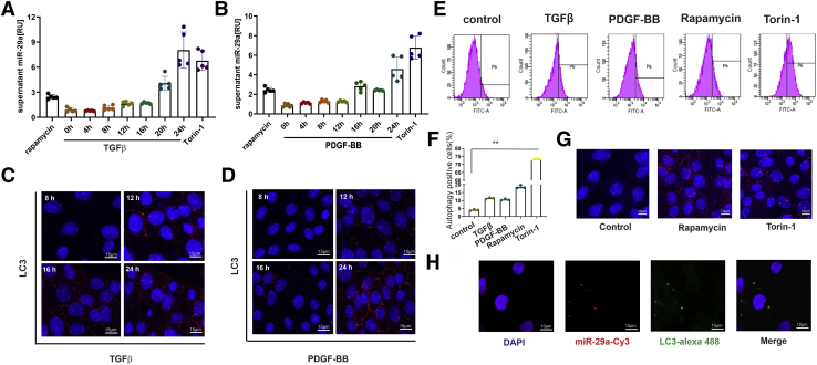
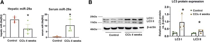
Intracellular and extracellular miRNA levels of primary and immortalized myofibroblastic HSC in response to profibrogenic stimulation by transforming growth factor β (TGFβ) or platelet-derived growth factor-BB (PDGF-BB) or upon inhibition of vesicular transport and autophagy processes were determined by quantitative polymerase chain reaction. Autophagy flux was studied by electron microscopy, flow cytometry, immunoblotting, and immunocytochemistry. Hepatic and serum miR-29a levels were quantified by using both liver tissue and serum samples from a cohort of chronic hepatitis C virus patients and a murine CCl induced liver fibrosis model.

RESULTS

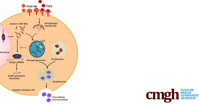
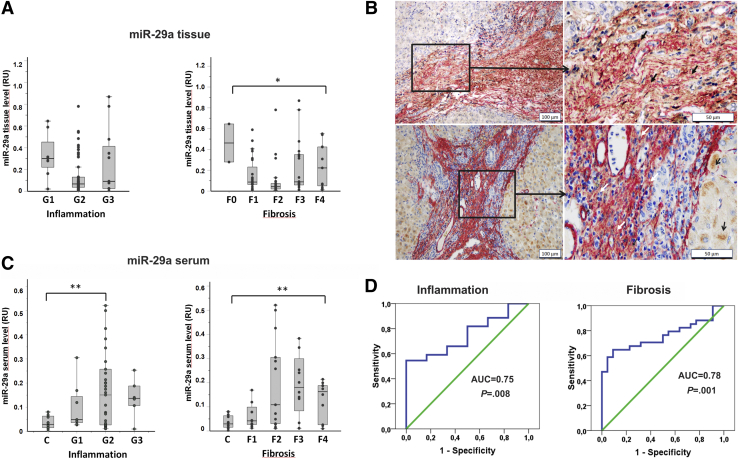
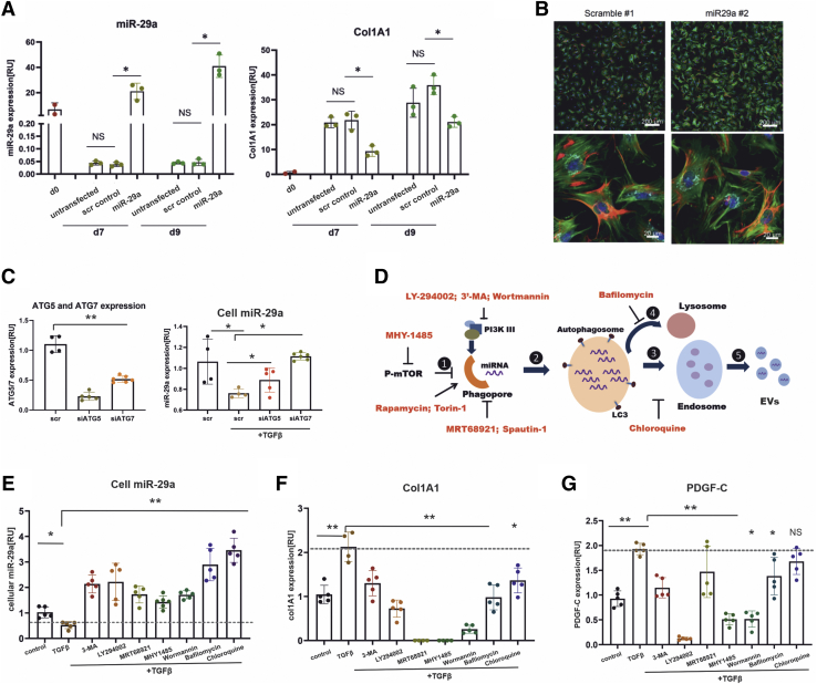
In our study, we show that TGFβ and PDGF-BB resulted in decrease of intracellular miR-29a and a pronounced increase of vesicular miR-29a release into the supernatant. Strikingly, miR-29a vesicular release was accompanied by enhanced autophagic activity and up-regulation of the autophagy marker protein LC3. Moreover, autophagy inhibition strongly prevented miR-29a secretion and repressed its targets' expression such as Col1A1. Consistently, hepatic miR-29a loss and increased LC3 expression in myofibroblastic HSC were associated with increased serum miR-29a levels in CCl-treated murine liver fibrosis and specimens of hepatitis C virus patients with chronic liver disease.

CONCLUSIONS

We provide evidence that activation-associated autophagy in HSC induces release of miR-29a, whereas inhibition of autophagy represses fibrogenic gene expression in part through attenuated miR-29a secretion.

摘要

背景与目的

肝纤维化由长期慢性肝损伤引起,伴有细胞外基质(ECM)间质积累的加速伤口愈合反应。活化的肝星状细胞(HSC)是 ECM 产生的主要来源。微小 RNA29a(miR-29a)是一种重要的抗纤维化 miRNA,在纤维化过程中受到抑制,导致胶原合成上调。

方法

通过定量聚合酶链反应测定转化生长因子β(TGFβ)或血小板衍生生长因子-BB(PDGF-BB)致纤维化刺激或抑制囊泡运输和自噬过程对原代和永生化肌成纤维细胞 HSC 的细胞内和细胞外 miRNA 水平。通过电子显微镜、流式细胞术、免疫印迹和免疫细胞化学研究自噬流。通过使用慢性丙型肝炎病毒患者的肝组织和血清样本以及 CCl 诱导的肝纤维化模型,定量测定肝和血清 miR-29a 水平。

结果

在我们的研究中,我们表明 TGFβ 和 PDGF-BB 导致细胞内 miR-29a 减少,并显著增加囊泡 miR-29a 释放到上清液中。引人注目的是,miR-29a 囊泡释放伴随着增强的自噬活性和自噬标志物蛋白 LC3 的上调。此外,自噬抑制强烈阻止 miR-29a 分泌并抑制其靶基因如 Col1A1 的表达。一致地,在 CCl 处理的肝纤维化小鼠模型和丙型肝炎病毒患者慢性肝病的标本中,肌成纤维细胞 HSC 中与激活相关的自噬导致 miR-29a 丢失和 LC3 表达增加,与血清 miR-29a 水平升高相关。

结论

我们提供的证据表明,HSC 中激活相关的自噬诱导 miR-29a 的释放,而自噬抑制通过减弱 miR-29a 的分泌部分抑制成纤维基因表达。

https://cdn.ncbi.nlm.nih.gov/pmc/blobs/6fde/9046234/cd48a5d05854/gr6.jpg
https://cdn.ncbi.nlm.nih.gov/pmc/blobs/6fde/9046234/3204f9acd6f7/fx1.jpg
https://cdn.ncbi.nlm.nih.gov/pmc/blobs/6fde/9046234/4357731cf563/gr1.jpg
https://cdn.ncbi.nlm.nih.gov/pmc/blobs/6fde/9046234/57f2283500c1/gr2.jpg
https://cdn.ncbi.nlm.nih.gov/pmc/blobs/6fde/9046234/09919612548e/gr3.jpg
https://cdn.ncbi.nlm.nih.gov/pmc/blobs/6fde/9046234/0d28c04080a1/gr4.jpg
https://cdn.ncbi.nlm.nih.gov/pmc/blobs/6fde/9046234/8231cfacf143/gr5.jpg
https://cdn.ncbi.nlm.nih.gov/pmc/blobs/6fde/9046234/cd48a5d05854/gr6.jpg

相似文献

1
Autophagy-Related Activation of Hepatic Stellate Cells Reduces Cellular miR-29a by Promoting Its Vesicular Secretion.自噬相关的肝星状细胞激活通过促进其囊泡分泌来减少细胞 miR-29a。
Cell Mol Gastroenterol Hepatol. 2022;13(6):1701-1716. doi: 10.1016/j.jcmgh.2022.02.013. Epub 2022 Feb 24.
2
Hepatocyte growth factor (HGF) inhibits collagen I and IV synthesis in hepatic stellate cells by miRNA-29 induction.肝细胞生长因子(HGF)通过 miRNA-29 的诱导抑制肝星状细胞中胶原 I 和 IV 的合成。
PLoS One. 2011;6(9):e24568. doi: 10.1371/journal.pone.0024568. Epub 2011 Sep 9.
3
Activation of Mir-29a in Activated Hepatic Stellate Cells Modulates Its Profibrogenic Phenotype through Inhibition of Histone Deacetylases 4.活化肝星状细胞中Mir-29a的激活通过抑制组蛋白脱乙酰基酶4调节其促纤维化表型。
PLoS One. 2015 Aug 25;10(8):e0136453. doi: 10.1371/journal.pone.0136453. eCollection 2015.
4
MicroRNA-29a Alleviates Bile Duct Ligation Exacerbation of Hepatic Fibrosis in Mice through Epigenetic Control of Methyltransferases.微小RNA-29a通过对甲基转移酶的表观遗传调控减轻小鼠胆管结扎所致的肝纤维化恶化
Int J Mol Sci. 2017 Jan 18;18(1):192. doi: 10.3390/ijms18010192.
5
Hepatic stellate cell autophagy inhibits extracellular vesicle release to attenuate liver fibrosis.肝星状细胞自噬抑制细胞外囊泡释放以减轻肝纤维化。
J Hepatol. 2020 Nov;73(5):1144-1154. doi: 10.1016/j.jhep.2020.04.044. Epub 2020 May 8.
6
miR-29a-3p suppresses hepatic fibrosis pathogenesis by modulating hepatic stellate cell proliferation via targeting PIK3R3 gene expression.miR-29a-3p 通过靶向 PIK3R3 基因表达抑制肝星状细胞增殖从而抑制肝纤维化发病机制。
Biochem Biophys Res Commun. 2020 Sep 3;529(4):922-929. doi: 10.1016/j.bbrc.2020.06.102. Epub 2020 Jul 30.
7
Exogenous Therapeutics of Microrna-29a Attenuates Development of Hepatic Fibrosis in Cholestatic Animal Model through Regulation of Phosphoinositide 3-Kinase p85 Alpha.外源性 miR-29a 治疗通过调节磷酸肌醇 3-激酶 p85α 减轻胆汁淤积动物模型肝纤维化的发展。
Int J Mol Sci. 2020 May 21;21(10):3636. doi: 10.3390/ijms21103636.
8
Systemic Delivery of scAAV8-Encoded MiR-29a Ameliorates Hepatic Fibrosis in Carbon Tetrachloride-Treated Mice.经系统递送编码miR-29a的scAAV8可改善四氯化碳处理小鼠的肝纤维化。
PLoS One. 2015 Apr 29;10(4):e0124411. doi: 10.1371/journal.pone.0124411. eCollection 2015.
9
MicroRNA-30a ameliorates hepatic fibrosis by inhibiting Beclin1-mediated autophagy.微小 RNA-30a 通过抑制 Beclin1 介导的自噬来改善肝纤维化。
J Cell Mol Med. 2017 Dec;21(12):3679-3692. doi: 10.1111/jcmm.13278. Epub 2017 Aug 1.
10
MicroRNA-503 Targets Mothers Against Decapentaplegic Homolog 7 Enhancing Hepatic Stellate Cell Activation and Hepatic Fibrosis.miRNA-503 靶向抑制 Decapentaplegic 同源物 7 增强肝星状细胞激活和肝纤维化。
Dig Dis Sci. 2021 Jun;66(6):1928-1939. doi: 10.1007/s10620-020-06460-7. Epub 2020 Jul 9.

引用本文的文献

1
Extracellular Vesicles and Micro-RNAs in Liver Disease.肝病中的细胞外囊泡与微小RNA
Semin Liver Dis. 2025 Jun;45(2):167-179. doi: 10.1055/a-2494-2233. Epub 2024 Dec 3.
2
N-Acetylcysteine Inhibits Coxsackievirus B3 Replication by Downregulating Eukaryotic Translation Elongation Factor 1 Alpha 1.N-乙酰半胱氨酸通过下调真核翻译延伸因子 1α1 抑制柯萨奇病毒 B3 复制。
Viruses. 2024 Sep 23;16(9):1503. doi: 10.3390/v16091503.
3
The Role of Epigenetic Mechanisms in the Pathogenesis of Hepatitis C Infection.表观遗传机制在丙型肝炎感染发病机制中的作用。

本文引用的文献

1
Hepatic stellate cell autophagy inhibits extracellular vesicle release to attenuate liver fibrosis.肝星状细胞自噬抑制细胞外囊泡释放以减轻肝纤维化。
J Hepatol. 2020 Nov;73(5):1144-1154. doi: 10.1016/j.jhep.2020.04.044. Epub 2020 May 8.
2
Lysosomal Exocytosis, Exosome Release and Secretory Autophagy: The Autophagic- and Endo-Lysosomal Systems Go Extracellular.溶酶体胞吐作用、外泌体释放和分泌自噬:自噬和内溶酶体系统向细胞外延伸。
Int J Mol Sci. 2020 Apr 8;21(7):2576. doi: 10.3390/ijms21072576.
3
Ursodeoxycholic acid alleviates experimental liver fibrosis involving inhibition of autophagy.
Biomolecules. 2024 Aug 10;14(8):986. doi: 10.3390/biom14080986.
4
Liver Fibrosis: From Basic Science towards Clinical Progress, Focusing on the Central Role of Hepatic Stellate Cells.肝纤维化:从基础科学到临床进展,聚焦于肝星状细胞的核心作用。
Int J Mol Sci. 2024 Jul 18;25(14):7873. doi: 10.3390/ijms25147873.
5
Naringenin alleviates liver fibrosis by triggering autophagy-dependent ferroptosis in hepatic stellate cells.柚皮素通过触发肝星状细胞中自噬依赖性铁死亡来减轻肝纤维化。
Heliyon. 2024 Mar 29;10(7):e28865. doi: 10.1016/j.heliyon.2024.e28865. eCollection 2024 Apr 15.
6
Cell-type specific role of autophagy in the liver and its implications in non-alcoholic fatty liver disease.自噬在肝脏中的细胞类型特异性作用及其在非酒精性脂肪性肝病中的意义。
World J Hepatol. 2023 Dec 27;15(12):1272-1283. doi: 10.4254/wjh.v15.i12.1272.
7
Found in translation-Fibrosis in metabolic dysfunction-associated steatohepatitis (MASH).翻译发现——代谢功能障碍相关脂肪性肝炎(MASH)中的纤维化
Sci Transl Med. 2023 Oct 4;15(716):eadi0759. doi: 10.1126/scitranslmed.adi0759.
8
Promotion Effect of Coexposure to a High-Fat Diet and Nano-Diethylnitrosamine on the Progression of Fatty Liver Malignant Transformation into Liver Cancer.高脂饮食与纳米二乙基亚硝胺共同暴露对脂肪肝恶性转化为肝癌进程的促进作用。
Int J Mol Sci. 2023 Sep 15;24(18):14162. doi: 10.3390/ijms241814162.
9
MicroRNA-29a Mitigates Laminectomy-Induced Spinal Epidural Fibrosis and Gait Dysregulation by Repressing TGF-β1 and IL-6.微小 RNA-29a 通过抑制 TGF-β1 和 IL-6 减轻椎板切除术后脊髓硬膜外纤维化和步态失调。
Int J Mol Sci. 2023 May 23;24(11):9158. doi: 10.3390/ijms24119158.
10
Circulating miRNA-192 and miR-29a as Disease Progression Biomarkers in Hepatitis C Patients with a Prevalence of HCV Genotype 3.循环 miRNA-192 和 miR-29a 作为丙型肝炎患者 HCV 基因 3 型流行中的疾病进展生物标志物。
Genes (Basel). 2023 May 8;14(5):1056. doi: 10.3390/genes14051056.
熊去氧胆酸通过抑制自噬缓解实验性肝纤维化。
Life Sci. 2020 Feb 1;242:117175. doi: 10.1016/j.lfs.2019.117175. Epub 2019 Dec 13.
4
TGF-β in Hepatic Stellate Cell Activation and Liver Fibrogenesis-Updated 2019.TGF-β 在肝星状细胞激活和肝纤维化中的作用-2019 更新版。
Cells. 2019 Nov 11;8(11):1419. doi: 10.3390/cells8111419.
5
IGFBPrP1 accelerates autophagy and activation of hepatic stellate cells via mutual regulation between H19 and PI3K/AKT/mTOR pathway.IGFBPrP1 通过 H19 与 PI3K/AKT/mTOR 通路的相互调节加速自噬和肝星状细胞的激活。
Biomed Pharmacother. 2019 Aug;116:109034. doi: 10.1016/j.biopha.2019.109034. Epub 2019 May 29.
6
Minimal information for studies of extracellular vesicles 2018 (MISEV2018): a position statement of the International Society for Extracellular Vesicles and update of the MISEV2014 guidelines.细胞外囊泡研究的最低限度信息2018(MISEV2018):国际细胞外囊泡协会的立场声明及MISEV2014指南的更新
J Extracell Vesicles. 2018 Nov 23;7(1):1535750. doi: 10.1080/20013078.2018.1535750. eCollection 2018.
7
Relevance of Autophagy in Parenchymal and Non-Parenchymal Liver Cells for Health and Disease.自噬在实质和非实质肝细胞中的相关性及其在健康和疾病中的作用。
Cells. 2019 Jan 1;8(1):16. doi: 10.3390/cells8010016.
8
Artificial MicroRNA-Mediated Tgfbr2 and Pdgfrb Co-Silencing Ameliorates Carbon Tetrachloride-Induced Hepatic Fibrosis in Mice.人工 microRNA 介导的 TGFBR2 和 PDGFRB 共沉默改善四氯化碳诱导的小鼠肝纤维化。
Hum Gene Ther. 2019 Feb;30(2):179-196. doi: 10.1089/hum.2018.047. Epub 2018 Nov 13.
9
Chloroquine inhibits autophagic flux by decreasing autophagosome-lysosome fusion.氯喹通过减少自噬体-溶酶体融合来抑制自噬流。
Autophagy. 2018;14(8):1435-1455. doi: 10.1080/15548627.2018.1474314. Epub 2018 Jul 20.
10
The many-faced role of autophagy in liver diseases.自噬在肝脏疾病中的多面作用。
J Hepatol. 2018 Mar;68(3):593-594. doi: 10.1016/j.jhep.2017.09.015. Epub 2018 Jan 17.