Grings Andreas, Jobic Camille, Kuwert Torsten, Ritt Philipp
Clinic of Nuclear Medicine, University Hospital Erlangen, Erlangen, Germany.
EJNMMI Phys. 2022 Mar 14;9(1):18. doi: 10.1186/s40658-022-00446-2.
Single-photon emission computed tomography (SPECT) can cause an over- or underestimation of tissue activity concentration due to limitations in spatial resolution compared to the structures under study. This is commonly referred to as partial volume effect (PVE). Ideally, the PVE should be controlled for and corrected. One such correction method involves determining recovery coefficients (RC) from phantom measurements. In the literature, several studies applying simplified geometries are available. In this study, we aimed to determine kidney PVE for realistic kidney geometries. Furthermore, we proposed a new surrogate metric for predicting the extent of PVE in kidneys.
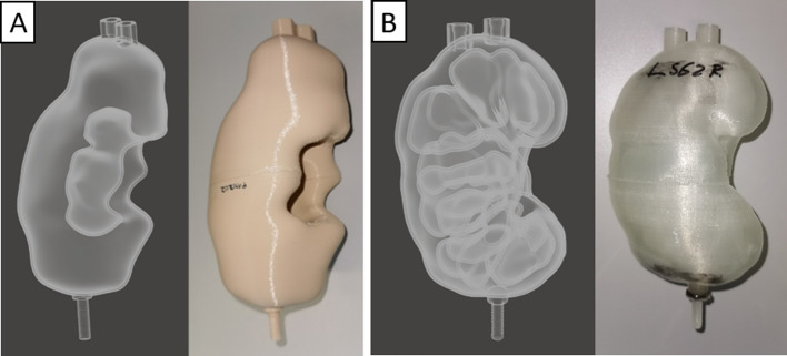
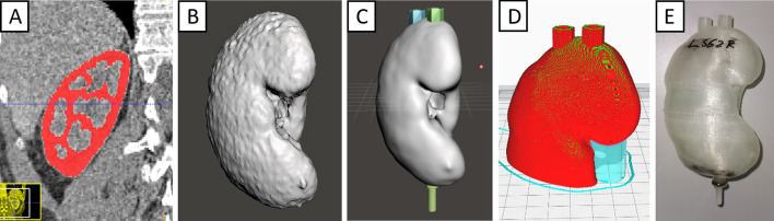
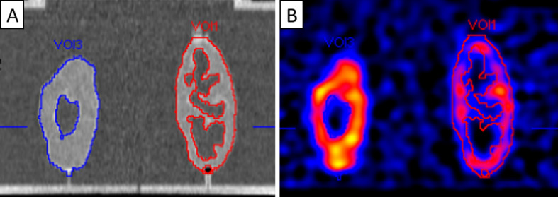
Based on patients' CT data, we manufactured fillable phantoms using a 3D-printer. Nine cortex-only and ten whole-parenchyma phantoms were obtained, and one ellipsoidal phantom for comparison. To measure PVE, we placed the phantoms in a torso phantom and filled them with a specified activity concentration. The phantoms' RCs were determined from fully quantitative SPECT/CT acquisitions at three different target-to-background ratios (TBRs). Additionally, the surface area-to-volume (SA:V) ratio was determined for all phantoms and correlated with RCs.
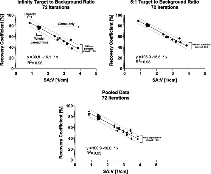
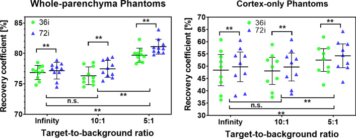
For SPECT reconstructions with 36 iterations, average RC ± one standard deviation at a 10-to-1 TBR was 76.3 ± 1.5% and 48.4 ± 8.3% for whole-parenchyma and cortex-only phantoms, respectively. The RC for the ellipsoidal phantom was 85.4%. The RC for whole-parenchyma was significantly higher than for cortex-only phantoms (p < 0.01). The RC variance was significantly higher for cortex-only phantoms (p < 0.01). A highly significant correlation of the SA:V ratio and RC was found for all phantoms. (R of linear regression was between 0.96 and 0.98.) CONCLUSION: Changes in the specific shape of the kidneys cause large changes in PVE magnitude. Therefore, RCs derived from more simple phantoms are most likely insufficient to correct the PVE in patient images. Furthermore, one should account for the fact that intra-renal activity distribution significantly influences the extent of PVE. Additionally, we found that the SA:V ratio excellently models kidney RCs; potentially, this approach could also be applied to other geometries and represents an alternative to full imaging process simulations to determine the extent of PVE.
与所研究的结构相比,单光子发射计算机断层扫描(SPECT)由于空间分辨率的限制,可能会导致组织活性浓度的高估或低估。这通常被称为部分容积效应(PVE)。理想情况下,应该对PVE进行控制和校正。一种校正方法是通过体模测量确定恢复系数(RC)。在文献中,有几项研究采用了简化的几何形状。在本研究中,我们旨在确定实际肾脏几何形状下的肾脏PVE。此外,我们提出了一种新的替代指标来预测肾脏中PVE的程度。
基于患者的CT数据,我们使用3D打印机制作了可填充的体模。获得了9个仅含皮质的体模和10个全实质体模,以及1个用于比较的椭球体模。为了测量PVE,我们将体模放置在躯干体模中,并用指定的活性浓度填充它们。通过在三种不同的靶本底比(TBR)下进行完全定量的SPECT/CT采集来确定体模的RC。此外,确定了所有体模的表面积与体积(SA:V)比,并将其与RC相关联。
对于进行36次迭代的SPECT重建,在10:1的TBR下,全实质体模和仅含皮质体模的平均RC±一个标准差分别为76.3± 1.5%和48.4± 8.3%。椭球体模的RC为85.4%。全实质体模的RC显著高于仅含皮质的体模(p < 0.01)。仅含皮质体模的RC方差显著更高(p < 0.01)。发现所有体模的SA:V比与RC高度显著相关。(线性回归的R在0.96至0.98之间。)结论:肾脏特定形状的变化会导致PVE大小的巨大变化。因此,从更简单的体模得出的RC很可能不足以校正患者图像中的PVE。此外,应该考虑到肾内活性分布会显著影响PVE的程度。此外,我们发现SA:V比能很好地模拟肾脏的RC;潜在地,这种方法也可以应用于其他几何形状,并且是确定PVE程度的全成像过程模拟的一种替代方法。