Laboratory of Reproductive Biology, University Hospital of Copenhagen, Rigshospitalet, Copenhagen, Denmark.
Department of Clinical Medicine, University of Copenhagen, Copenhagen, Denmark.
Front Endocrinol (Lausanne). 2022 Mar 10;13:853482. doi: 10.3389/fendo.2022.853482. eCollection 2022.
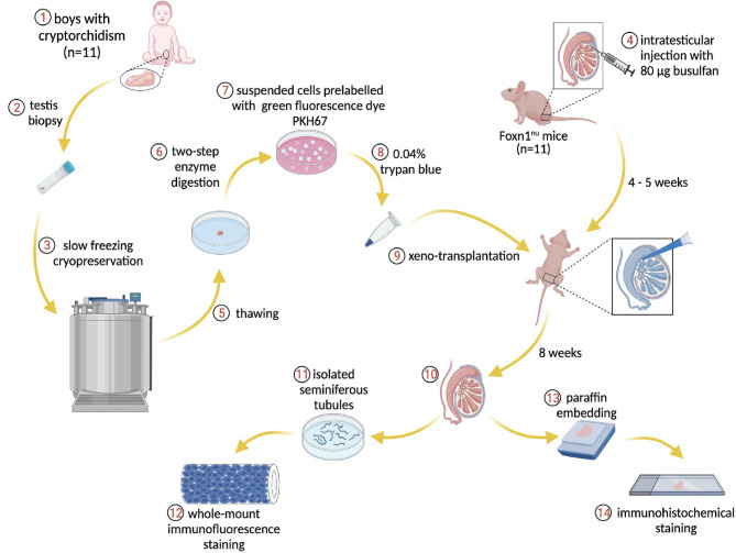
Cryopreservation of prepubertal testicular tissue preserves spermatogonial stem cells (SSCs) that may be used to restore fertility in men at risk of infertility due to gonadotoxic treatments for either a malignant or non-malignant disease. Spermatogonial stem cell-based transplantation is a promising fertility restoration technique. Previously, we performed xenotransplantation of propagated SSCs from prepubertal testis and found human SSCs colonies within the recipient testes six weeks post-transplantation. In order to avoid the propagation step of SSCs that may cause genetic and epigenetic changes, we performed direct injection of single cell suspension in this study, which potentially may be safer and easier to be applied in future clinical applications.
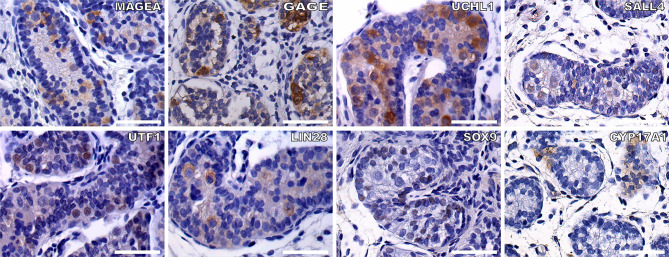
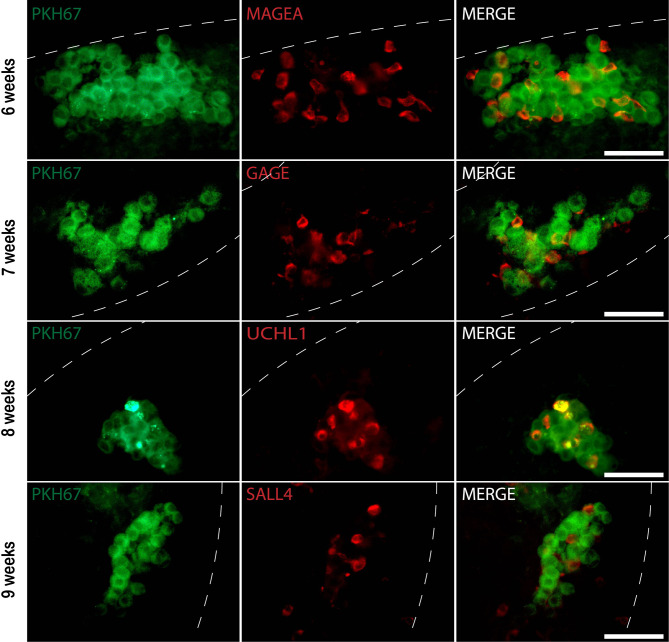
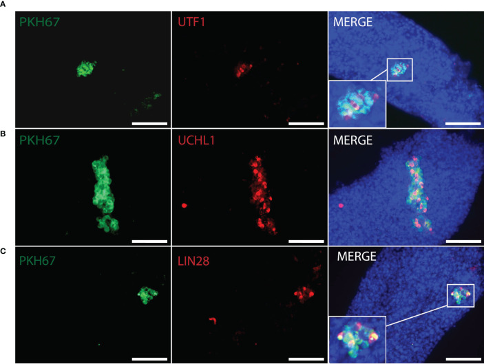
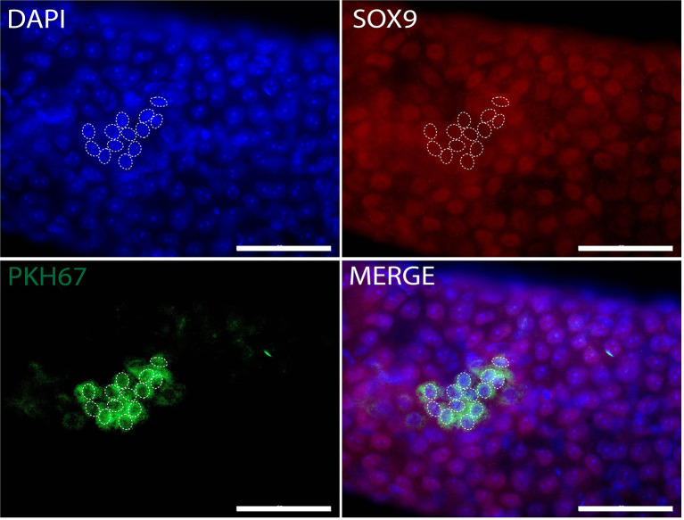
Testis biopsies were obtained from 11 infant boys (median age 1.3 years, range 0.5-3.5) with cryptorchidism. Following enzymatic digestion, dissociated single-cell suspensions were prelabeled with green fluorescent dye and directly transplanted into seminiferous tubules of busulfan-treated mice. Six to nine weeks post-transplantation, the presence of gonocytes and SSCs was determined by whole-mount immunofluorescence for a number of germ cell markers (MAGEA, GAGE, UCHL1, SALL4, UTF1, and LIN28), somatic cell markers (SOX9, CYP17A1).
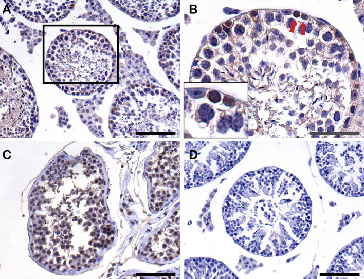
Following xenotransplantation human infant germ cells, consisting of gonocytes and SSCs, were shown to settle on the basal membrane of the recipient seminiferous tubules and form SSC colonies with expression of MAGEA, GAGE, UCHL1, SALL4, UTF1, and LIN28. The colonization efficiency was approximately 6%. No human Sertoli cells were detected in the recipient mouse testes.
Xenotransplantation, without propagation, of testicular cell suspensions from infant boys with cryptorchidism resulted in colonization of mouse seminiferous tubules six to nine weeks post-transplantation. Spermatogonial stem cell-based transplantation could be a therapeutic treatment for infertility of prepubertal boys with cryptorchidism and boys diagnosed with cancer. However, more studies are required to investigate whether the low number of the transplanted SSC is sufficient to secure the presence of sperm in the ejaculate of those patients over time.
通过对青春期前睾丸组织的冷冻保存,可以保留精原干细胞(SSC),这些干细胞可用于因恶性或非恶性疾病的性腺毒性治疗而面临生育能力受损风险的男性恢复生育能力。基于精原干细胞的移植是一种很有前途的生育力恢复技术。此前,我们进行了青春期前睾丸来源的增殖性 SSC 的异种移植,并在移植后 6 周于受体睾丸中发现了人 SSC 集落。为了避免 SSC 增殖可能导致遗传和表观遗传改变,我们在本研究中直接注射单细胞悬液,这可能更安全,并且更容易在未来的临床应用中应用。
从 11 名患有隐睾症的婴儿男孩(中位年龄 1.3 岁,范围 0.5-3.5 岁)中获取睾丸活检。在酶消化后,分离的单细胞悬液用绿色荧光染料预先标记,然后直接移植到白消安处理的小鼠的生精小管中。移植后 6-9 周,通过针对多种生殖细胞标志物(MAGEA、GAGE、UCHL1、SALL4、UTF1 和 LIN28)和体细胞标志物(SOX9、CYP17A1)的全器官免疫荧光检测来确定是否存在精原细胞和 SSC。
异种移植人婴儿生殖细胞后,精原细胞和 SSC 定居在受体生精小管的基底膜上,并形成表达 MAGEA、GAGE、UCHL1、SALL4、UTF1 和 LIN28 的 SSC 集落。定植效率约为 6%。在受体小鼠睾丸中未检测到人支持细胞。
来自隐睾症婴儿的睾丸细胞悬液未经增殖的异种移植,在移植后 6-9 周导致小鼠生精小管的定植。基于精原干细胞的移植可能是治疗青春期前隐睾症男孩和诊断为癌症男孩不育的一种治疗方法。然而,需要更多的研究来研究移植的 SSC 数量是否足以确保这些患者在一段时间后精液中存在精子。