Park Haewook, Han Kook Nam, Choi Byeong Hyeon, Yoon Hyunsuk, An Hyun Joon, Lee Jae Sung, Kim Hyun Koo
Department of Biomedical Sciences, Seoul National University College of Medicine, Seoul, Republic of Korea.
Department of Nuclear Medicine, Seoul National University College of Medicine, Seoul, Republic of Korea.
Transl Lung Cancer Res. 2022 Apr;11(4):588-599. doi: 10.21037/tlcr-21-909.
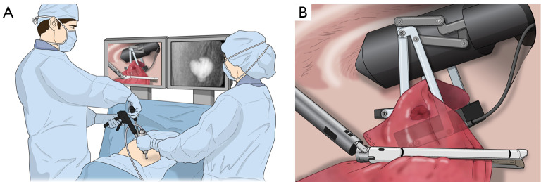
With advances in surgical technology, thoracic surgeons have widely adopted minimally invasive limited-resection techniques to preserve normal tissues. However, it remains difficult to achieve localization of invisible pulmonary nodules during surgery. Therefore, we proposed an ultra-low-dose X-ray imaging device for intraoperative pulmonary nodule localization during minimally invasive surgeries.
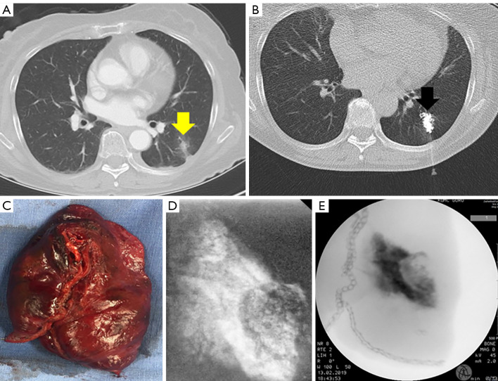
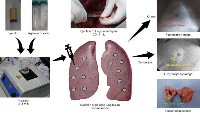
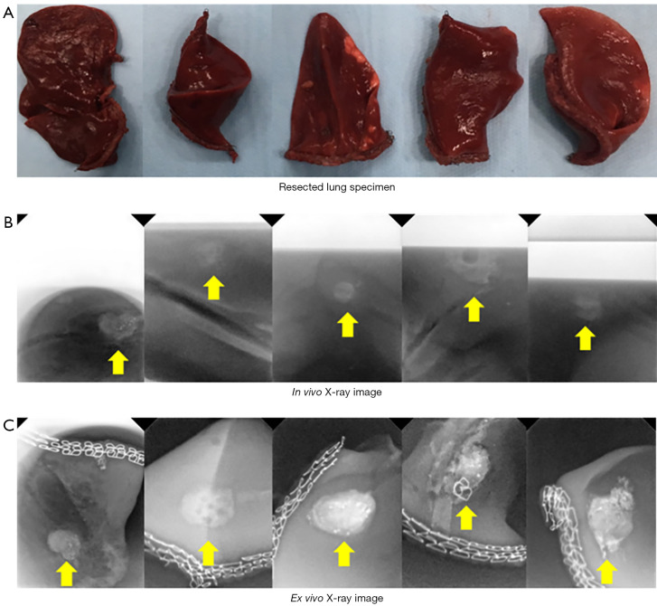
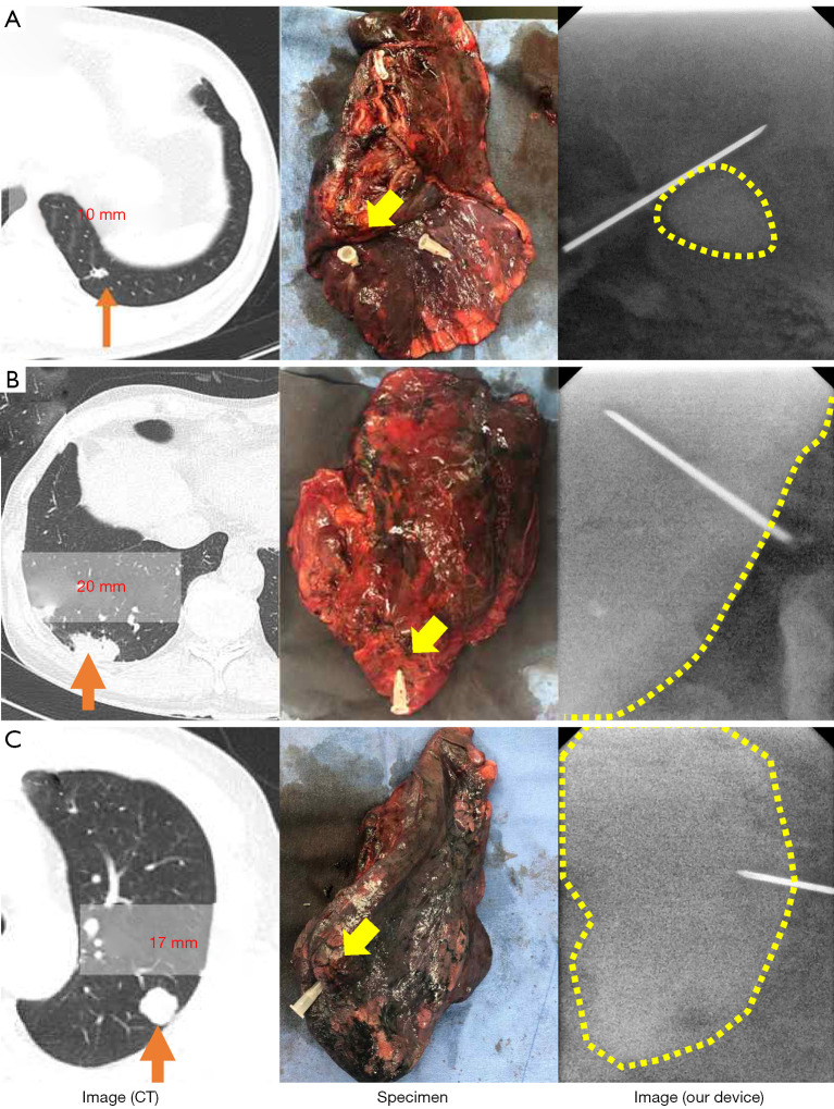
The proposed device features a hand-held type and consists of a carbon nanotube-based X-ray source and an intraoral dental sensor. In a preclinical study, we created pseudo pulmonary nodules using pig lungs. Subsequently, its clinical feasibility was evaluated using lung cancer specimens from patients with cancer who had undergone minimally invasive surgery.
Using the proposed device, we successfully differentiated normal and abnormal tissues from X-ray images of resected lung specimens. In addition, our proposed device only yielded an average radiation dose of 90.9 nGy for a single acquisition of X-ray images and demonstrated excellent temperature stability under consecutive X-ray irradiations. The radiation exposure of our proposed device (0.1±0.0006 µSv/h) was significantly lower than that of conventional C-arm fluoroscopy (41.5±51.8 µSv/h). In both preclinical and clinical studies, the margin of nodule shadows was clearly visualized using the proposed device.
The proposed device substantially reduced radiation exposure to staff and patients and may allow localization of pulmonary nodules. Our proposed device clearly revealed the margins of lung nodules with radiocontrast injection and showed the potential to identify solid nodules without the use of radiocontrast agents.
随着手术技术的进步,胸外科医生广泛采用微创有限切除技术以保留正常组织。然而,在手术过程中实现对不可见肺结节的定位仍然困难。因此,我们提出了一种用于微创手术中肺结节术中定位的超低剂量X射线成像设备。
所提出的设备为手持式,由基于碳纳米管的X射线源和口腔内牙科传感器组成。在一项临床前研究中,我们使用猪肺制造了假肺结节。随后,使用接受过微创手术的癌症患者的肺癌标本评估其临床可行性。
使用所提出的设备,我们成功地从切除的肺标本的X射线图像中区分出正常组织和异常组织。此外,我们提出的设备单次采集X射线图像的平均辐射剂量仅为90.9纳戈瑞,并且在连续X射线照射下表现出优异的温度稳定性。我们提出的设备的辐射暴露(0.1±0.0006微希沃特/小时)明显低于传统C形臂荧光透视(41.5±51.8微希沃特/小时)。在临床前和临床研究中,使用所提出的设备均能清晰地看到结节阴影的边缘。
所提出的设备大幅降低了对工作人员和患者的辐射暴露,并且可能实现肺结节的定位。我们提出的设备通过注射造影剂清晰地显示了肺结节的边缘,并显示出在不使用造影剂的情况下识别实性结节的潜力。