Department of Clinical and Experimental Epilepsy, UCL Queen Square Institute of Neurology, Box 29, Queen Square, London, WC1N 3BG, UK.
Chalfont Centre for Epilepsy, Chalfont St Peter, Bucks, SL9 0RJ, UK.
Acta Neuropathol. 2022 Jul;144(1):107-127. doi: 10.1007/s00401-022-02429-0. Epub 2022 May 12.
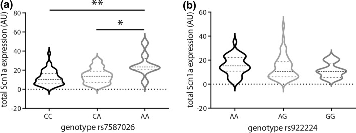
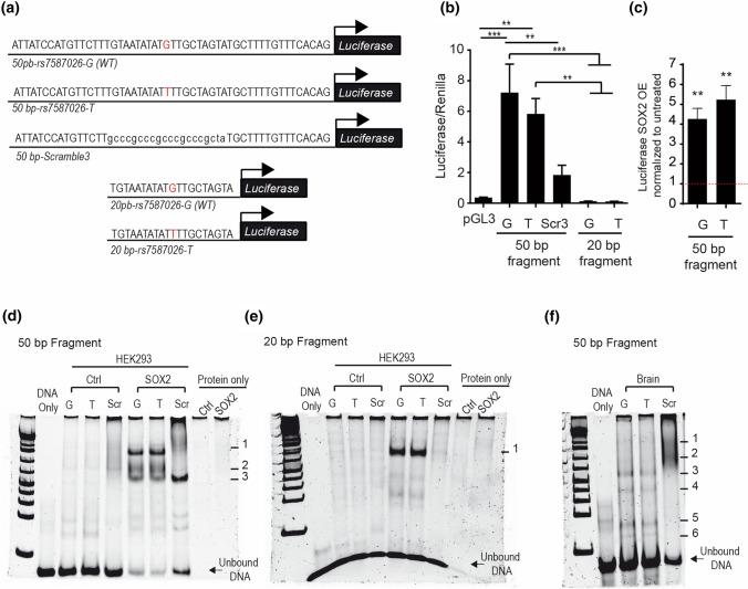
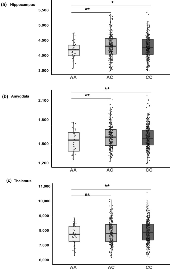
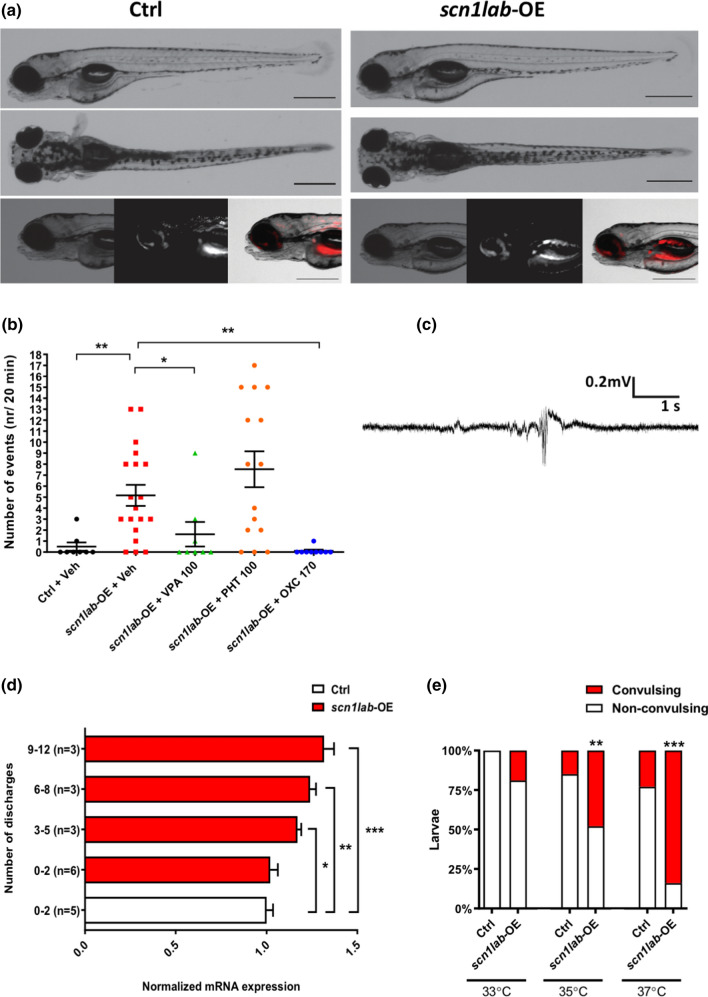
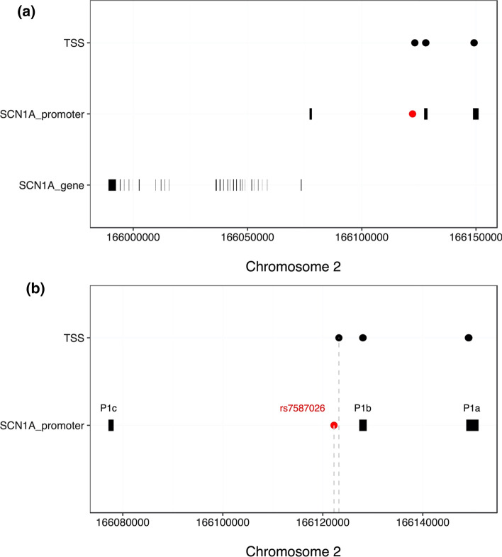
Mesial temporal lobe epilepsy with hippocampal sclerosis and a history of febrile seizures is associated with common variation at rs7587026, located in the promoter region of SCN1A. We sought to explore possible underlying mechanisms. SCN1A expression was analysed in hippocampal biopsy specimens of individuals with mesial temporal lobe epilepsy with hippocampal sclerosis who underwent surgical treatment, and hippocampal neuronal cell loss was quantitatively assessed using immunohistochemistry. In healthy individuals, hippocampal volume was measured using MRI. Analyses were performed stratified by rs7587026 type. To study the functional consequences of increased SCN1A expression, we generated, using transposon-mediated bacterial artificial chromosome transgenesis, a zebrafish line expressing exogenous scn1a, and performed EEG analysis on larval optic tecta at 4 day post-fertilization. Finally, we used an in vitro promoter analysis to study whether the genetic motif containing rs7587026 influences promoter activity. Hippocampal SCN1A expression differed by rs7587026 genotype (Kruskal-Wallis test P = 0.004). Individuals homozygous for the minor allele showed significantly increased expression compared to those homozygous for the major allele (Dunn's test P = 0.003), and to heterozygotes (Dunn's test P = 0.035). No statistically significant differences in hippocampal neuronal cell loss were observed between the three genotypes. Among 597 healthy participants, individuals homozygous for the minor allele at rs7587026 displayed significantly reduced mean hippocampal volume compared to major allele homozygotes (Cohen's D = - 0.28, P = 0.02), and to heterozygotes (Cohen's D = - 0.36, P = 0.009). Compared to wild type, scn1lab-overexpressing zebrafish larvae exhibited more frequent spontaneous seizures [one-way ANOVA F(4,54) = 6.95 (P < 0.001)]. The number of EEG discharges correlated with the level of scn1lab overexpression [one-way ANOVA F(4,15) = 10.75 (P < 0.001]. Finally, we showed that a 50 bp promoter motif containing rs7587026 exerts a strong regulatory role on SCN1A expression, though we could not directly link this to rs7587026 itself. Our results develop the mechanistic link between rs7587026 and mesial temporal lobe epilepsy with hippocampal sclerosis and a history of febrile seizures. Furthermore, we propose that quantitative precision may be important when increasing SCN1A expression in current strategies aiming to treat seizures in conditions involving SCN1A haploinsufficiency, such as Dravet syndrome.
内侧颞叶癫痫伴海马硬化和热性惊厥史与 rs7587026 常见变异有关,该变异位于 SCN1A 启动子区域。我们试图探索潜在的机制。对接受手术治疗的内侧颞叶癫痫伴海马硬化患者的海马活检标本进行 SCN1A 表达分析,并通过免疫组织化学定量评估海马神经元细胞丢失。在健康个体中,使用 MRI 测量海马体积。按 rs7587026 类型进行分层分析。为了研究 SCN1A 表达增加的功能后果,我们使用转座子介导的细菌人工染色体转基因技术生成了表达外源性 scn1a 的斑马鱼系,并在受精后 4 天对幼虫的光顶盖进行 EEG 分析。最后,我们使用体外启动子分析来研究包含 rs7587026 的遗传基序是否影响启动子活性。SCN1A 在海马中的表达因 rs7587026 基因型而异(Kruskal-Wallis 检验 P=0.004)。与主要等位基因纯合子相比,携带次要等位基因的个体表现出明显增加的表达(Dunn 检验 P=0.003),并且与杂合子相比(Dunn 检验 P=0.035)。三种基因型之间未观察到海马神经元细胞丢失的统计学差异。在 597 名健康参与者中,与 rs7587026 中的主要等位基因纯合子相比,携带次要等位基因的个体的平均海马体积明显减小(Cohen's D=-0.28,P=0.02),并且与杂合子相比(Cohen's D=-0.36,P=0.009)。与野生型相比,scn1lab 过表达斑马鱼幼虫表现出更频繁的自发性癫痫发作[单因素方差分析 F(4,54)=6.95(P<0.001)]。EEG 放电次数与 scn1lab 过表达水平相关[单因素方差分析 F(4,15)=10.75(P<0.001)]。最后,我们表明包含 rs7587026 的 50bp 启动子基序对 SCN1A 表达具有很强的调节作用,但我们不能直接将其与 rs7587026 本身联系起来。我们的结果在机制上连接了 rs7587026 与内侧颞叶癫痫伴海马硬化和热性惊厥史。此外,我们提出在当前旨在治疗涉及 SCN1A 杂合不足的情况下发生的癫痫发作的策略中增加 SCN1A 表达时,定量精度可能很重要,例如 Dravet 综合征。