Ochiai Jun, Villanueva Larakaye, Niihara Hope, Niihara Yutaka, Oliva Joan
Emmaus Life Sciences, Inc., Torrance, CA, United States.
Front Cell Dev Biol. 2022 Apr 26;10:873603. doi: 10.3389/fcell.2022.873603. eCollection 2022.
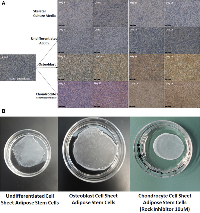
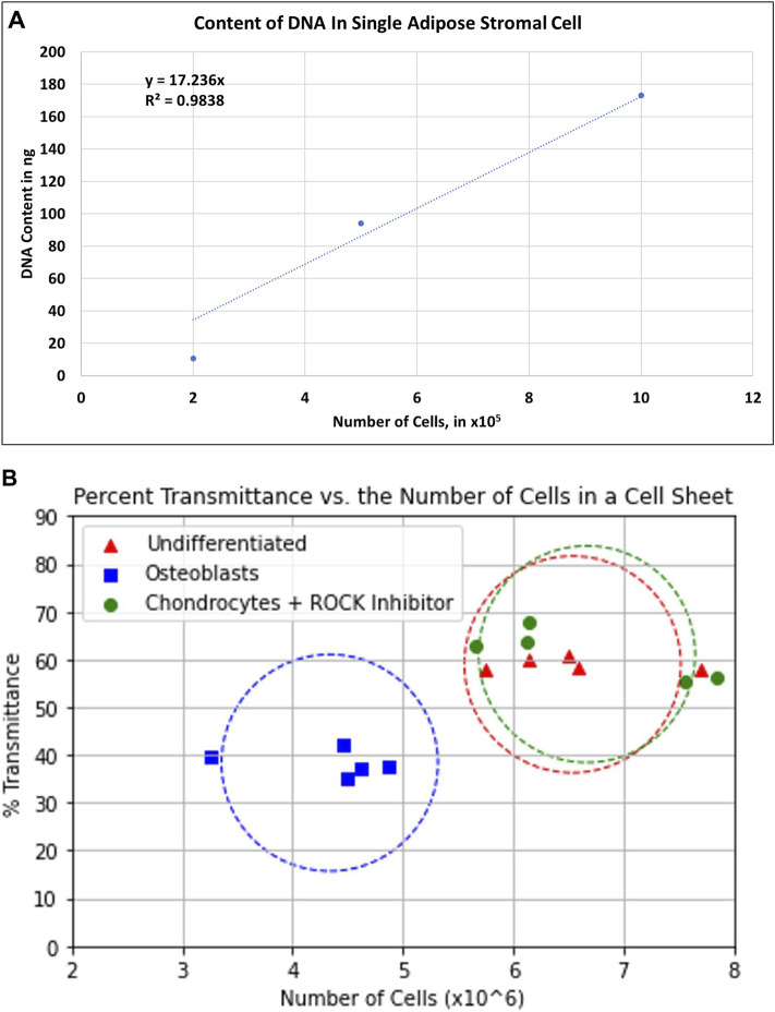
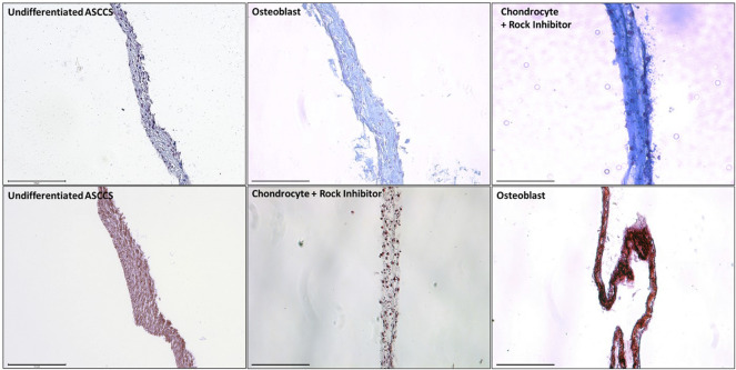
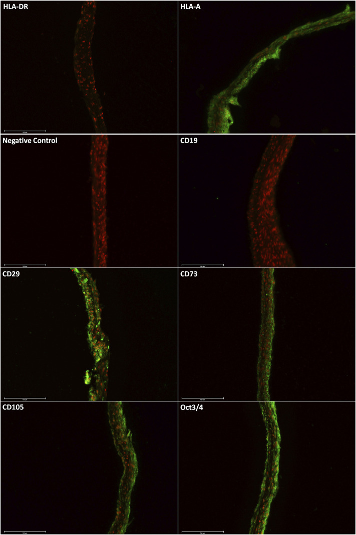
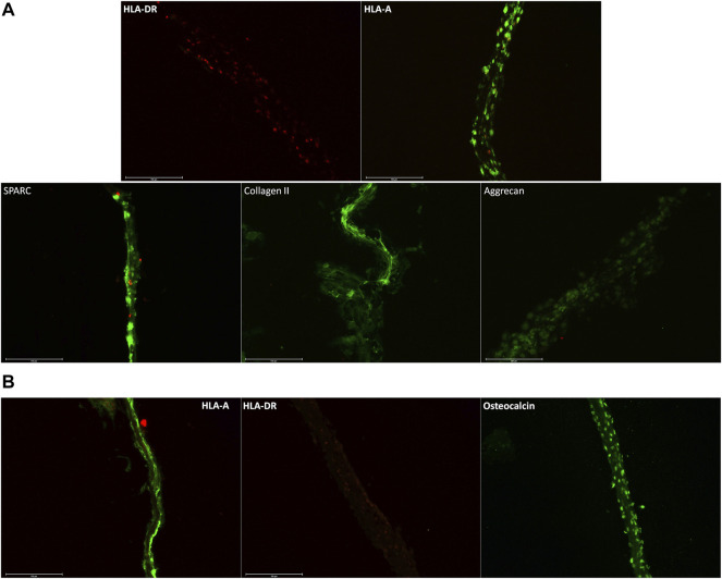
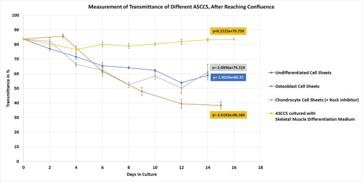
Well-characterized adipose stem cells and chemically defined culture media are important factors that control the production of the cell sheet, used in translational medicine. In this study, we have developed and engineered multilayer adipose stem cell cell sheets (ASCCSs) using chemically defined/serum-free culture media: undifferentiated or differentiated into osteoblasts and chondrocytes. In addition, using the cell sheet transmittance, we estimated the number of cells per cell sheet. Undifferentiated ASCCSs were engineered in 10 days, using serum-free/xeno-free culture media. They were CD29, CD73, CD90, CD105+, HLA-A+, and HLA-DR-. ASCCSs differentiated into chondrocytes and osteoblasts were also engineered using chemically defined and animal-free culture media, in only 14 days. The addition of an ROCK inhibitor improved the chondrocyte cell sheet engineering. The decrease in the cell sheet transmittance rate was higher for the osteoblast cell sheets due to the intracellular Ca accumulation. The estimation of cell number per cell sheet was carried out with the transmittance, which will provide important information for cell sheet posology. In conclusion, three types of ASCCSs were engineered using serum-free, xeno-free culture media, expressing their specific markers. Their transmittance measurement allowed estimating the number of cells per cell sheet, with a non-invasive methodology.
特征明确的脂肪干细胞和化学成分明确的培养基是控制用于转化医学的细胞片生产的重要因素。在本研究中,我们使用化学成分明确/无血清培养基开发并构建了多层脂肪干细胞片(ASCCSs):未分化的或分化为成骨细胞和软骨细胞的。此外,我们利用细胞片的透光率估算了每张细胞片中的细胞数量。未分化的ASCCSs在无血清/无异种成分培养基中培养10天即可构建完成。它们表达CD29、CD73、CD90、CD105+、HLA-A+,且不表达HLA-DR-。分化为软骨细胞和成骨细胞的ASCCSs同样使用化学成分明确且无动物成分的培养基,仅需14天即可构建完成。添加ROCK抑制剂可改善软骨细胞片的构建。由于细胞内钙的积累,成骨细胞片的细胞片透光率下降幅度更大。通过透光率对每张细胞片中的细胞数量进行估算,这将为细胞片给药方案提供重要信息。总之,我们使用无血清、无异种成分的培养基构建了三种类型的ASCCSs,它们表达各自的特异性标志物。通过对其透光率的测量,我们能够采用非侵入性方法估算每张细胞片中的细胞数量。