Department of Radiology, University of California San Diego, 200 W. Arbor Drive, San Diego, CA 92103, United States.
Department of Radiology, University of California San Diego, 200 W. Arbor Drive, San Diego, CA 92103, United States; Center for Functional Magnetic Resonance Imaging, University of California San Diego, 9500 Gilman Drive, La Jolla, CA 92093, United States.
Neuroimage Clin. 2022;35:103017. doi: 10.1016/j.nicl.2022.103017. Epub 2022 Apr 30.
The pathophysiologic basis of posterior reversible encephalopathy syndrome (PRES) remains controversial. Hypertension (HTN)-induced autoregulatory failure with subsequent hyperperfusion is the leading hypothesis, whereas alternative theories suggest vasoconstriction-induced hypoperfusion as the underlying mechanism. Studies using contrast-based CT and MR perfusion imaging have yielded contradictory results supporting both ideas. This work represents one of the first applications of arterial spin labeling (ASL) to evaluate cerebral blood flow (CBF) changes in PRES.
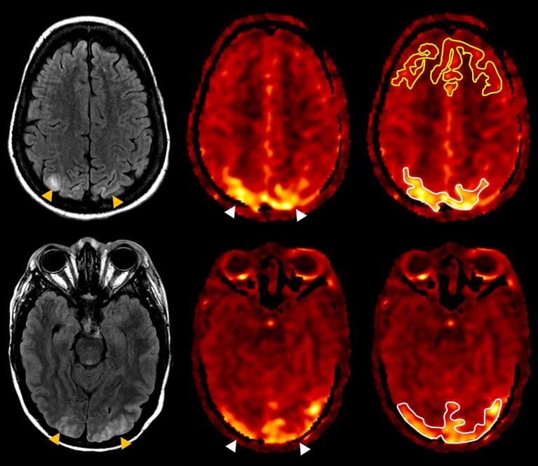
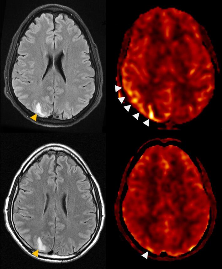
After obtaining Institutional Review Board approval, MRI reports at our institution from 07/2015 to 09/2020 were retrospectively searched and reviewed for mention of "PRES" and "posterior reversible encephalopathy syndrome." Of the resulting 103 MRIs (performed on GE 1.5 Tesla or 3 Tesla scanners), 20 MRIs in 18 patients who met the inclusion criteria of clinical and imaging diagnosis of PRES and had diagnostic-quality pseudocontinuous ASL scans were included. Patients with a more likely alternative diagnosis, technically non-diagnostic ASL, or other intracranial abnormalities limiting assessment of underlying PRES features were excluded. Perfusion in FLAIR-affected brain regions was qualitatively assessed using ASL and characterized as hyperperfusion, normal, or hypoperfusion. Additional quantitative analysis was performed by measuring average gray matter CBF in abnormal versus normal brain regions.
HTN was the most common PRES etiology (65%). ASL showed hyperperfusion in 13 cases and normal perfusion in 7 cases. A hypoperfusion pattern was not identified. Quantitative analysis of gray matter CBF among patients with visually apparent hyperperfusion showed statistically higher perfusion in affected versus normal appearing brain regions (median CBF 100.4 ml/100 g-min vs. 61.0 ml/ 100 g-min, p < 0.001).
Elevated ASL CBF was seen in the majority (65%) of patients with PRES, favoring the autoregulatory failure hypothesis as a predominant mechanism. Our data support ASL as a practical way to assess and noninvasively monitor cerebral perfusion in PRES that could potentially alter management strategies.
后部可逆性脑病综合征(PRES)的病理生理基础仍存在争议。高血压(HTN)诱导的自动调节失败伴随后续的过度灌注是主要假说,而替代理论则认为是血管收缩诱导的低灌注是潜在的机制。使用基于对比的 CT 和 MR 灌注成像的研究得出了支持这两种观点的相互矛盾的结果。这项工作代表了动脉自旋标记(ASL)首次应用于评估 PRES 中的脑血流(CBF)变化。
在获得机构审查委员会批准后,我们对本机构 2015 年 7 月至 2020 年 9 月的 MRI 报告进行了回顾性搜索和审查,以查找“PRES”和“后部可逆性脑病综合征”的提及。在总共 103 份 MRI(在 GE 1.5 Tesla 或 3 Tesla 扫描仪上进行)中,有 20 份 MRI 来自 18 名符合 PRES 的临床和影像学诊断标准且具有诊断质量的假性连续 ASL 扫描的患者,这些患者被纳入研究。患有更可能的替代诊断、技术上非诊断性 ASL 或其他限制评估潜在 PRES 特征的颅内异常的患者被排除在外。使用 ASL 对 FLAIR 受影响的脑区的灌注进行定性评估,并将其特征描述为过度灌注、正常或低灌注。通过测量异常与正常脑区之间的平均灰质 CBF 进行了额外的定量分析。
HTN 是 PRES 最常见的病因(65%)。ASL 显示 13 例过度灌注,7 例正常灌注。未发现低灌注模式。对视觉上明显过度灌注的患者进行灰质 CBF 的定量分析显示,在受影响的脑区与正常脑区相比,灌注明显更高(中位 CBF 100.4 ml/100 g-min 与 61.0 ml/100 g-min,p<0.001)。
在大多数 PRES 患者(65%)中,ASL 的 CBF 升高,这有利于自动调节失败假说作为主要机制。我们的数据支持 ASL 作为一种实用的方法来评估和无创监测 PRES 中的脑灌注,这可能会改变管理策略。