Department of Health Laboratory Technology, School of Public Health, China Medical University, Shenyang 110122, China.
Key Laboratory of Arsenic-Related Biological Effects and Prevention and Treatment in Liaoning Province, School of Public Health, China Medical University, Shenyang 110122, China.
Int J Mol Sci. 2022 May 19;23(10):5697. doi: 10.3390/ijms23105697.
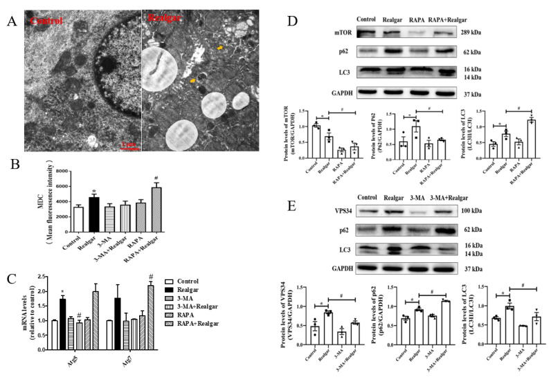
Realgar, a poisonous traditional Chinese medicine, has been shown to cause liver injury when used for long periods or overdoses. However, the underlying molecular mechanisms and therapeutic targets have not been fully elucidated. The aim of this study is to explore the role of autophagy in sub-chronic realgar exposure-induced liver injury. Here, the liver injury model was established by continuously administrating mice with 1.35 g/kg realgar for 8 weeks. 3-methyladenine (3-MA) and rapamycin (RAPA) were used to regulate autophagy. The results showed that realgar induced abnormal changes in liver function, pathological morphology, expression of inflammatory cytokines, and upregulated NLRP3 inflammasome pathway in mouse livers. RAPA treatment (an inducer of autophagy) significantly improved realgar-induced liver injury and NLRP3 inflammasome activation, while 3-MA (an inhibitor of autophagy) aggravated the realgar-induced liver injury and NLRP3 inflammasome activation. Furthermore, we found that realgar-induced NLRP3 inflammasome activation in mouse livers is mediated by ROS. RAPA eliminates excessive ROS, inhibits NF-κB nuclear translocation and down-regulates the TXNIP/NLRP3 axis, consequently suppressing ROS-mediated NLRP3 inflammasome activation, which may be the underlying mechanism of the protective effect of autophagy on realgar-induced liver injury. In conclusion, the results of this study suggest that autophagy alleviates realgar-induced liver injury by inhibiting ROS-mediated NLRP3 inflammasome activation. Autophagy may represent a therapeutic target in modulating realgar-induced liver injury.
雄黄是一种传统的中药,长期或过量使用会导致肝损伤。然而,其潜在的分子机制和治疗靶点尚未完全阐明。本研究旨在探讨自噬在亚慢性雄黄暴露诱导的肝损伤中的作用。在此,通过连续 8 周给小鼠 1.35 g/kg 雄黄建立肝损伤模型。使用 3-甲基腺嘌呤(3-MA)和雷帕霉素(RAPA)调节自噬。结果表明,雄黄诱导小鼠肝脏功能异常、病理形态改变、炎症细胞因子表达上调及 NLRP3 炎性小体通路激活。RAPA 处理(自噬诱导剂)显著改善了雄黄诱导的肝损伤和 NLRP3 炎性小体激活,而 3-MA(自噬抑制剂)则加重了雄黄诱导的肝损伤和 NLRP3 炎性小体激活。此外,我们发现雄黄诱导的小鼠肝脏 NLRP3 炎性小体激活是由 ROS 介导的。RAPA 消除了过量的 ROS,抑制了 NF-κB 核易位,并下调了 TXNIP/NLRP3 轴,从而抑制了 ROS 介导的 NLRP3 炎性小体激活,这可能是自噬对雄黄诱导的肝损伤的保护作用的潜在机制。总之,本研究结果表明,自噬通过抑制 ROS 介导的 NLRP3 炎性小体激活来减轻雄黄诱导的肝损伤。自噬可能成为调节雄黄诱导的肝损伤的治疗靶点。