• 文献检索
  • 文档翻译
  • 深度研究
  • 学术资讯
  • Suppr Zotero 插件Zotero 插件
  • 邀请有礼
  • 套餐&价格
  • 历史记录
应用&插件
Suppr Zotero 插件Zotero 插件浏览器插件Mac 客户端Windows 客户端微信小程序
定价
高级版会员购买积分包购买API积分包
服务
文献检索文档翻译深度研究API 文档MCP 服务
关于我们
关于 Suppr公司介绍联系我们用户协议隐私条款
关注我们

Suppr 超能文献

核心技术专利:CN118964589B侵权必究
粤ICP备2023148730 号-1Suppr @ 2026

文献检索

告别复杂PubMed语法,用中文像聊天一样搜索,搜遍4000万医学文献。AI智能推荐,让科研检索更轻松。

立即免费搜索

文件翻译

保留排版,准确专业,支持PDF/Word/PPT等文件格式,支持 12+语言互译。

免费翻译文档

深度研究

AI帮你快速写综述,25分钟生成高质量综述,智能提取关键信息,辅助科研写作。

立即免费体验

鉴定 SARS-CoV-2 膜蛋白(M)与增殖细胞核抗原(PCNA)之间的相互作用,将其作为潜在的治疗靶点。

Characterization of the Interaction Between SARS-CoV-2 Membrane Protein (M) and Proliferating Cell Nuclear Antigen (PCNA) as a Potential Therapeutic Target.

机构信息

Multidisciplinary Laboratory of Food and Health, School of Applied Sciences, University of Campinas (Unicamp), Limeira, Brazil.

Laboratory of Signaling Mechanisms, School of Pharmaceutical Sciences, University of Campinas, (Unicamp), Campinas, Brazil.

出版信息

Front Cell Infect Microbiol. 2022 May 23;12:849017. doi: 10.3389/fcimb.2022.849017. eCollection 2022.

DOI:10.3389/fcimb.2022.849017
PMID:35677658
原文链接:https://pmc.ncbi.nlm.nih.gov/articles/PMC9168989/
Abstract

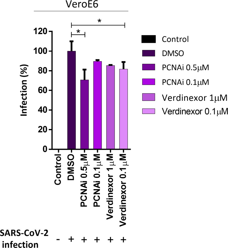
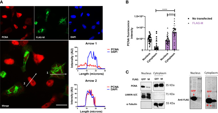
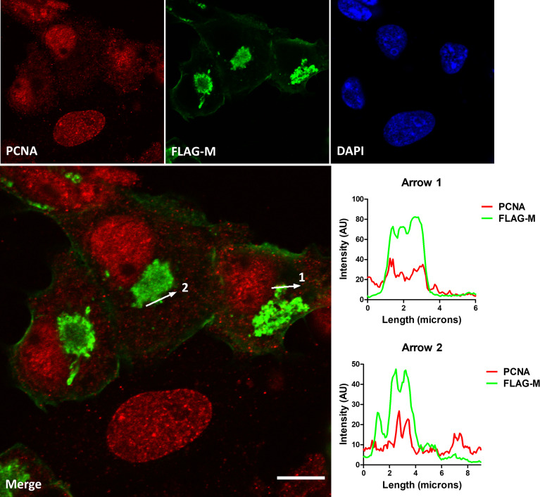
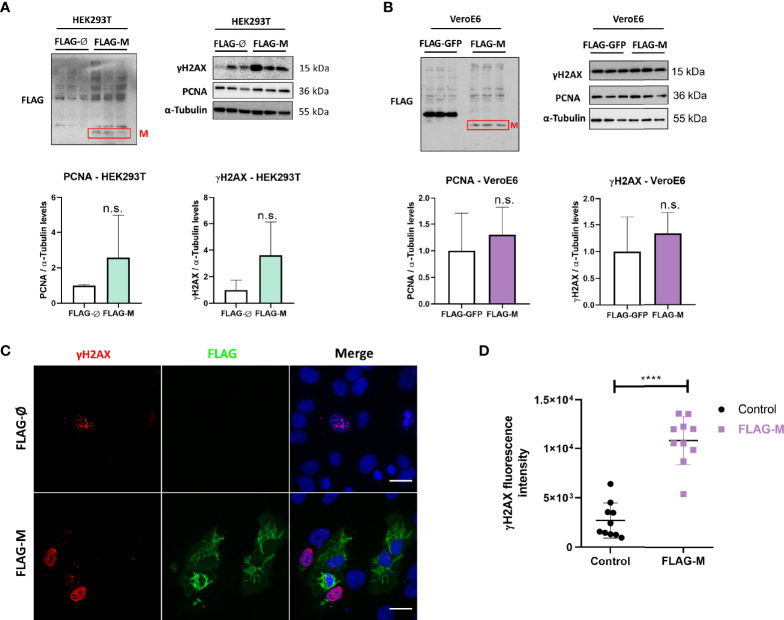
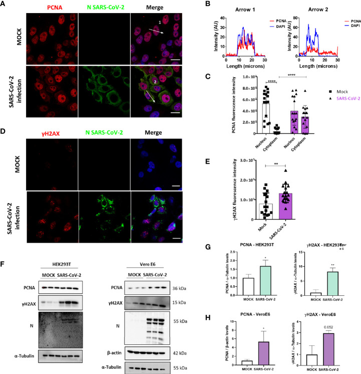
SARS-CoV-2 is an emerging virus from the Coronaviridae family and is responsible for the ongoing COVID-19 pandemic. In this work, we explored the previously reported SARS-CoV-2 structural membrane protein (M) interaction with human Proliferating Cell Nuclear Antigen (PCNA). The M protein is responsible for maintaining virion shape, and PCNA is a marker of DNA damage which is essential for DNA replication and repair. We validated the M-PCNA interaction through immunoprecipitation, immunofluorescence co-localization, and PLA (Proximity Ligation Assay). In cells infected with SARS-CoV-2 or transfected with M protein, using immunofluorescence and cell fractioning, we documented a reallocation of PCNA from the nucleus to the cytoplasm and the increase of PCNA and γH2AX (another DNA damage marker) expression. We also observed an increase in PCNA and γH2AX expression in the lung of a COVID-19 patient by immunohistochemistry. In addition, the inhibition of PCNA translocation by PCNA I1 and Verdinexor led to a reduction of plaque formation in an assay. We, therefore, propose that the transport of PCNA to the cytoplasm and its association with M could be a virus strategy to manipulate cell functions and may be considered a target for COVID-19 therapy.

摘要

SARS-CoV-2 是冠状病毒科的一种新兴病毒,是导致当前 COVID-19 大流行的罪魁祸首。在这项工作中,我们研究了先前报道的 SARS-CoV-2 结构膜蛋白(M)与人增殖细胞核抗原(PCNA)的相互作用。M 蛋白负责维持病毒粒子的形状,而 PCNA 是 DNA 损伤的标志物,对于 DNA 复制和修复至关重要。我们通过免疫沉淀、免疫荧光共定位和 PLA(邻近连接分析)验证了 M-PCNA 相互作用。在感染 SARS-CoV-2 或转染 M 蛋白的细胞中,通过免疫荧光和细胞分级分离,我们记录了 PCNA 从细胞核重新分配到细胞质,以及 PCNA 和 γH2AX(另一种 DNA 损伤标志物)表达的增加。我们还通过免疫组织化学观察到 COVID-19 患者肺组织中 PCNA 和 γH2AX 表达增加。此外,PCNA I1 和 Verdinexor 抑制 PCNA 易位导致斑块形成减少。因此,我们提出 PCNA 向细胞质的转运及其与 M 的结合可能是病毒操纵细胞功能的一种策略,可被视为 COVID-19 治疗的一个靶点。

https://cdn.ncbi.nlm.nih.gov/pmc/blobs/b5b2/9168989/e17fd42071d4/fcimb-12-849017-g006.jpg
https://cdn.ncbi.nlm.nih.gov/pmc/blobs/b5b2/9168989/04ff98b7abf9/fcimb-12-849017-g001.jpg
https://cdn.ncbi.nlm.nih.gov/pmc/blobs/b5b2/9168989/4bf1fe374a57/fcimb-12-849017-g002.jpg
https://cdn.ncbi.nlm.nih.gov/pmc/blobs/b5b2/9168989/7027918cf828/fcimb-12-849017-g003.jpg
https://cdn.ncbi.nlm.nih.gov/pmc/blobs/b5b2/9168989/fbc34849a530/fcimb-12-849017-g004.jpg
https://cdn.ncbi.nlm.nih.gov/pmc/blobs/b5b2/9168989/7f98d2429506/fcimb-12-849017-g005.jpg
https://cdn.ncbi.nlm.nih.gov/pmc/blobs/b5b2/9168989/e17fd42071d4/fcimb-12-849017-g006.jpg

相似文献

1
Characterization of the Interaction Between SARS-CoV-2 Membrane Protein (M) and Proliferating Cell Nuclear Antigen (PCNA) as a Potential Therapeutic Target.鉴定 SARS-CoV-2 膜蛋白(M)与增殖细胞核抗原(PCNA)之间的相互作用,将其作为潜在的治疗靶点。
Front Cell Infect Microbiol. 2022 May 23;12:849017. doi: 10.3389/fcimb.2022.849017. eCollection 2022.
2
Ubiquitin‑like protein FAT10 regulates DNA damage repair via modification of proliferating cell nuclear antigen.泛素样蛋白 FAT10 通过修饰增殖细胞核抗原调节 DNA 损伤修复。
Mol Med Rep. 2018 Jun;17(6):7487-7496. doi: 10.3892/mmr.2018.8843. Epub 2018 Apr 5.
3
The herpes simplex virus 1 UL36USP deubiquitinase suppresses DNA repair in host cells via deubiquitination of proliferating cell nuclear antigen.单纯疱疹病毒1型UL36USP去泛素化酶通过使增殖细胞核抗原去泛素化来抑制宿主细胞中的DNA修复。
J Biol Chem. 2017 May 19;292(20):8472-8483. doi: 10.1074/jbc.M117.778076. Epub 2017 Mar 27.
4
Nuclear insulin-like growth factor 1 receptor phosphorylates proliferating cell nuclear antigen and rescues stalled replication forks after DNA damage.细胞核胰岛素样生长因子1受体使增殖细胞核抗原磷酸化,并在DNA损伤后挽救停滞的复制叉。
J Biol Chem. 2017 Nov 3;292(44):18227-18239. doi: 10.1074/jbc.M117.781492. Epub 2017 Sep 18.
5
Proliferating Cell Nuclear Antigen Suppresses RNA Replication of through an Interaction with the Viral Genome.增殖细胞核抗原通过与病毒基因组相互作用抑制 的 RNA 复制。
J Virol. 2019 Oct 29;93(22). doi: 10.1128/JVI.00961-19. Print 2019 Nov 15.
6
Proliferating cell nuclear antigen inhibitors block distinct stages of herpes simplex virus infection.增殖细胞核抗原抑制剂阻断单纯疱疹病毒感染的不同阶段。
PLoS Pathog. 2023 Jul 24;19(7):e1011539. doi: 10.1371/journal.ppat.1011539. eCollection 2023 Jul.
7
Lys-110 is essential for targeting PCNA to replication and repair foci, and the K110A mutant activates apoptosis.赖氨酸-110对于将增殖细胞核抗原靶向至复制和修复位点至关重要,而K110A突变体可激活细胞凋亡。
Biol Cell. 2008 Dec;100(12):675-86. doi: 10.1042/BC20070158.
8
Analysis of proliferating cell nuclear antigen (PCNA) associated with DNA.与DNA相关的增殖细胞核抗原(PCNA)分析。
Methods Mol Biol. 2006;314:457-75. doi: 10.1385/1-59259-973-7:457.
9
The herpes simplex virus (HSV) protein ICP34.5 is a virion component that forms a DNA-binding complex with proliferating cell nuclear antigen and HSV replication proteins.单纯疱疹病毒(HSV)蛋白ICP34.5是一种病毒体成分,它与增殖细胞核抗原和HSV复制蛋白形成DNA结合复合物。
J Neurovirol. 2003 Aug;9(4):477-88. doi: 10.1080/13550280390218788.
10
Subcellular distribution of p21 and PCNA in normal and repair-deficient cells following DNA damage.DNA损伤后正常细胞和修复缺陷细胞中p21和增殖细胞核抗原(PCNA)的亚细胞分布。
Curr Biol. 1996 Feb 1;6(2):189-99. doi: 10.1016/s0960-9822(02)00452-9.

引用本文的文献

1
Unveiling the role of hexon-associated host proteins in fowl adenovirus serotype 4 replication.揭示六邻体相关宿主蛋白在4型禽腺病毒复制中的作用。
Front Vet Sci. 2025 Jun 3;12:1562872. doi: 10.3389/fvets.2025.1562872. eCollection 2025.
2
Structural proteins of human coronaviruses: what makes them different?人类冠状病毒的结构蛋白:它们有何不同之处?
Front Cell Infect Microbiol. 2024 Dec 6;14:1458383. doi: 10.3389/fcimb.2024.1458383. eCollection 2024.
3
Immune characteristics of kidney transplant recipients with acute respiratory distress syndrome induced by COVID-19 at single-cell resolution.

本文引用的文献

1
Selinexor, a novel selective inhibitor of nuclear export, reduces SARS-CoV-2 infection and protects the respiratory system in vivo.塞利尼索,一种新型核输出抑制剂,可降低 SARS-CoV-2 感染并保护体内呼吸系统。
Antiviral Res. 2021 Aug;192:105115. doi: 10.1016/j.antiviral.2021.105115. Epub 2021 Jun 19.
2
Multilevel proteomics reveals host perturbations by SARS-CoV-2 and SARS-CoV.多水平蛋白质组学揭示 SARS-CoV-2 和 SARS-CoV 对宿主的干扰。
Nature. 2021 Jun;594(7862):246-252. doi: 10.1038/s41586-021-03493-4. Epub 2021 Apr 12.
3
The SARS-CoV-2 nucleocapsid phosphoprotein forms mutually exclusive condensates with RNA and the membrane-associated M protein.
单细胞分辨率解析 COVID-19 诱导的急性呼吸窘迫综合征肾移植受者的免疫特征。
Respir Res. 2024 Jan 18;25(1):34. doi: 10.1186/s12931-024-02682-9.
4
Zebrafish models of COVID-19.COVID-19 的斑马鱼模型。
FEMS Microbiol Rev. 2023 Jan 16;47(1). doi: 10.1093/femsre/fuac042.
5
Computational Analysis of Short Linear Motifs in the Spike Protein of SARS-CoV-2 Variants Provides Possible Clues into the Immune Hijack and Evasion Mechanisms of Omicron Variant.计算分析 SARS-CoV-2 变异株刺突蛋白中的短线性基序,为奥密克戎变异株的免疫劫持和逃逸机制提供了可能的线索。
Int J Mol Sci. 2022 Aug 8;23(15):8822. doi: 10.3390/ijms23158822.
新型冠状病毒核衣壳磷蛋白与 RNA 和膜相关的 M 蛋白形成相互排斥的凝聚物。
Nat Commun. 2021 Jan 21;12(1):502. doi: 10.1038/s41467-020-20768-y.
4
Comparison of the SARS-CoV-2 (2019-nCoV) M protein with its counterparts of SARS-CoV and MERS-CoV species.严重急性呼吸综合征冠状病毒2(2019新型冠状病毒)M蛋白与其严重急性呼吸综合征冠状病毒和中东呼吸综合征冠状病毒对应蛋白的比较。
J King Saud Univ Sci. 2021 Mar;33(2):101335. doi: 10.1016/j.jksus.2020.101335. Epub 2021 Jan 7.
5
Severe acute respiratory syndrome coronavirus 2 (SARS-CoV-2) membrane (M) protein inhibits type I and III interferon production by targeting RIG-I/MDA-5 signaling.严重急性呼吸综合征冠状病毒 2(SARS-CoV-2)的膜(M)蛋白通过靶向 RIG-I/MDA-5 信号通路抑制 I 型和 III 型干扰素的产生。
Signal Transduct Target Ther. 2020 Dec 28;5(1):299. doi: 10.1038/s41392-020-00438-7.
6
SARS-CoV-2 structure and replication characterized by in situ cryo-electron tomography.利用原位冷冻电镜断层成像技术对 SARS-CoV-2 的结构和复制进行表征。
Nat Commun. 2020 Nov 18;11(1):5885. doi: 10.1038/s41467-020-19619-7.
7
A systemic and molecular study of subcellular localization of SARS-CoV-2 proteins.一项关于严重急性呼吸综合征冠状病毒2(SARS-CoV-2)蛋白亚细胞定位的系统和分子研究。
Signal Transduct Target Ther. 2020 Nov 17;5(1):269. doi: 10.1038/s41392-020-00372-8.
8
SARS-CoV-2 membrane glycoprotein M antagonizes the MAVS-mediated innate antiviral response.SARS-CoV-2 膜糖蛋白 M 拮抗 MAVS 介导的先天抗病毒反应。
Cell Mol Immunol. 2021 Mar;18(3):613-620. doi: 10.1038/s41423-020-00571-x. Epub 2020 Oct 27.
9
Virus-Host Interactome and Proteomic Survey Reveal Potential Virulence Factors Influencing SARS-CoV-2 Pathogenesis.病毒-宿主相互作用组和蛋白质组学调查揭示了影响 SARS-CoV-2 发病机制的潜在毒力因子。
Med. 2021 Jan 15;2(1):99-112.e7. doi: 10.1016/j.medj.2020.07.002. Epub 2020 Jul 21.
10
The Global Phosphorylation Landscape of SARS-CoV-2 Infection.新冠病毒感染的全球磷酸化组景观。
Cell. 2020 Aug 6;182(3):685-712.e19. doi: 10.1016/j.cell.2020.06.034. Epub 2020 Jun 28.