Granata Vincenza, Fusco Roberta, Belli Andrea, Danti Ginevra, Bicci Eleonora, Cutolo Carmen, Petrillo Antonella, Izzo Francesco
Division of Radiology, "Istituto Nazionale Tumori IRCCS Fondazione Pascale - IRCCS di Napoli", I-80131, Naples, Italy.
Medical Oncology Division, Igea Spa, Naples, Italy.
Infect Agent Cancer. 2022 Jun 9;17(1):25. doi: 10.1186/s13027-022-00441-3.
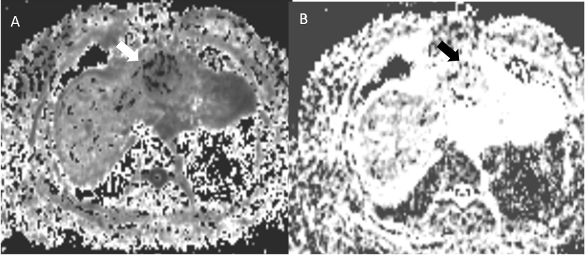
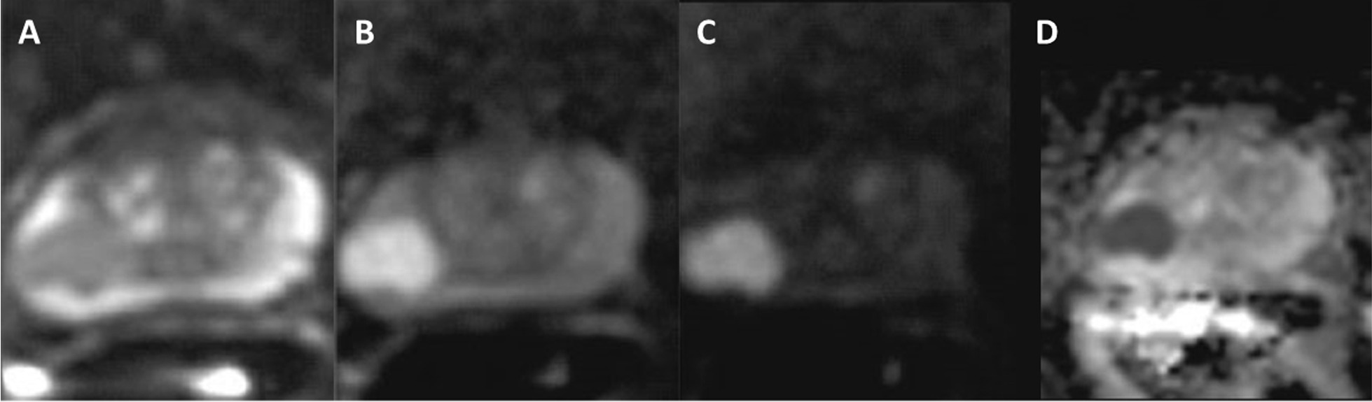
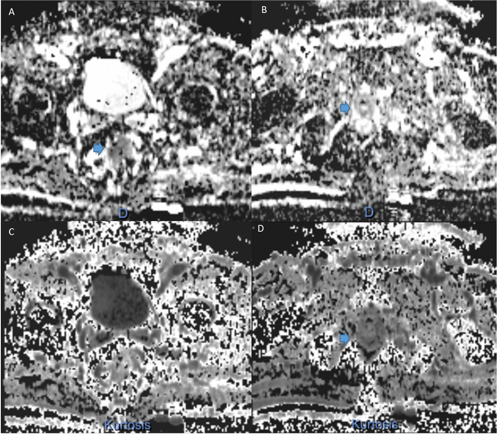
This article provides an overview of diffusion kurtosis (DKI) imaging in abdominal oncology. DKI allows for more data on tissue structures than the conventional diffusion model (DWI). However, DKI requires high quality images at b-values greater than 1000 s/mm and high signal-to-noise ratio (SNR) that traditionally MRI systems are not able to acquire and therefore there are generally amplified anatomical distortions on the images due to less homogeneity of the field. Advances in both hardware and software on modern MRI scanners have currently enabled ultra-high b-value imaging and offered the ability to apply DKI to multiple extracranial sites. Previous studies have evaluated the ability of DKI to characterize and discriminate tumor grade compared to conventional DWI. Additionally, in several studies the DKI sequences used were based on planar echo (EPI) acquisition, which is susceptible to motion, metal and air artefacts and prone to low SNRs and distortions, leading to low quality images for some small lesions, which may affect the accuracy of the results. Another problem is the optimal b-value of DKI, which remains to be explored and not yet standardized, as well as the manual selection of the ROI, which could affect the accuracy of some parameters.
本文概述了扩散峰度成像(DKI)在腹部肿瘤学中的应用。与传统扩散模型(DWI)相比,DKI能够提供更多关于组织结构的数据。然而,DKI需要在b值大于1000 s/mm²时获得高质量图像以及高信噪比(SNR),而传统MRI系统无法获取这些数据,因此由于磁场均匀性较差,图像上通常会出现放大的解剖结构扭曲。现代MRI扫描仪在硬件和软件方面的进步目前已实现超高b值成像,并能够将DKI应用于多个颅外部位。以往的研究评估了与传统DWI相比,DKI在表征和区分肿瘤分级方面的能力。此外,在一些研究中使用的DKI序列基于平面回波(EPI)采集,这种采集方式易受运动、金属和空气伪影的影响,且容易出现低信噪比和扭曲,导致一些小病变的图像质量较低,这可能会影响结果的准确性。另一个问题是DKI的最佳b值,这仍有待探索且尚未标准化,以及感兴趣区域(ROI)的手动选择,这可能会影响某些参数的准确性。