Department of Anesthesiology and Pain Medicine, Kangbuk Samsung Hospital, Sungkyunkwan University School of Medicine, Seoul, Korea.
Department of Anesthesiology and Pain Medicine, College of Medicine, The Catholic University of Korea, Seoul, Korea.
Korean J Anesthesiol. 2022 Dec;75(6):496-501. doi: 10.4097/kja.22153. Epub 2022 Jun 15.
Previous studies have demonstrated that morphological changes in the suprascapular notch are closely associated with suprascapular nerve entrapment syndrome (SNES). Thus, we hypothesized that the suprascapular notch cross-sectional area (SSNCSA) could be a good diagnostic parameter to assess SNES.
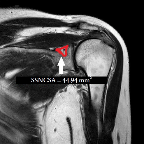
We acquired suprascapular notch data from 10 patients with SNES and 10 healthy individuals who had undergone shoulder magnetic resonance imaging (S-MRI) and had no evidence of SNES. T2-weighted coronal magnetic resonance images were acquired from the shoulder. We analyzed the SSNCSA at the shoulder on S-MRI using our image-analysis program (INFINITT PACS). The SSNCSA was measured as the suprascapular notch, which was the most affected site in coronal S-MRI images.
The mean SSNCSA was 64.50 ± 8.93 mm2 in the control group and 44.94 ± 10.40 mm2 in the SNES group. Patients with SNES had significantly lower SSNCSA (P < 0.01) than those in the control group. Receiver operating curve analysis showed that the best cut-off of the SSNCSA was 57.49 mm2, with 80.0% sensitivity, 80.0% specificity, and an area under the curve of 0.92 (95% CI [0.79, 1.00]).
The SSNCSA was found to have acceptable diagnostic properties for detecting SNES. We hope that these results will help diagnose SNES objectively.
先前的研究表明,肩胛上切迹的形态变化与肩胛上神经卡压综合征(SNES)密切相关。因此,我们假设肩胛上切迹横截面积(SSNCSA)可以作为评估 SNES 的良好诊断参数。
我们从 10 例 SNES 患者和 10 例健康个体中获得肩胛上切迹数据,这些个体均接受了肩部磁共振成像(S-MRI)检查,且没有 SNES 的证据。从肩部获取 T2 加权冠状磁共振图像。我们使用图像分析程序(INFINITT PACS)分析 S-MRI 上的 SSNCSA。SSNCSA 在冠状 S-MRI 图像中最受影响的肩胛上切迹处进行测量。
对照组的平均 SSNCSA 为 64.50±8.93mm2,SNES 组为 44.94±10.40mm2。SNES 患者的 SSNCSA 明显低于对照组(P<0.01)。受试者工作特征曲线分析显示,SSNCSA 的最佳截断值为 57.49mm2,具有 80.0%的敏感性、80.0%的特异性和 0.92 的曲线下面积(95%置信区间 [0.79, 1.00])。
SSNCSA 被发现具有可接受的诊断 SNES 的特性。我们希望这些结果将有助于客观地诊断 SNES。