Department of Medico-Surgical Sciences and Biotechnologies, Sapienza University of Rome Polo Pontino - I.C.O.T., Via Franco Faggiana 1668, 04100, Latina, Italy.
Section of Neurology, Department of Medicine and Surgery, University of Perugia, Perugia, Italy.
J Headache Pain. 2022 Jul 12;23(1):80. doi: 10.1186/s10194-022-01446-4.
Several functional neuroimaging studies on healthy controls and patients with migraine with aura have shown that the activation of functional networks during visual stimulation is not restricted to the striate system, but also includes several extrastriate networks.
Before and after 4 min of visual stimulation with a checkerboard pattern, we collected functional MRI in 21 migraine with aura (MwA) patients and 18 healthy subjects (HS). For each recording session, we identified independent resting-state networks in each group and correlated network connection strength changes with clinical disease features.
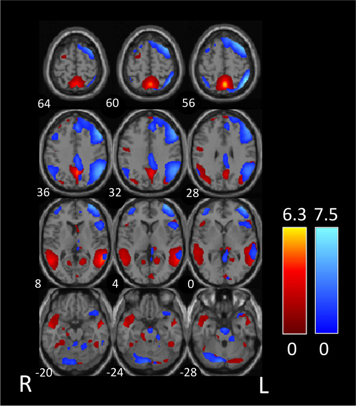
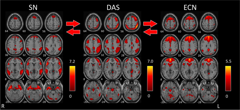
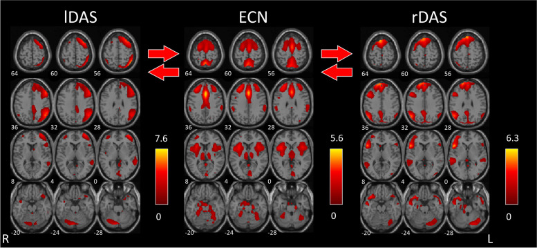
Before visual stimulation, we found reduced connectivity between the default mode network and the left dorsal attention system (DAS) in MwA patients compared to HS. In HS, visual stimulation increases functional connectivity between the independent components of the bilateral DAS and the executive control network (ECN). In MwA, visual stimulation significantly improved functional connectivity between the independent component pairs salience network and DAS, and between DAS and ECN. The ECN Z-scores after visual stimulation were negatively related to the monthly frequency of aura.
In individuals with MwA, 4 min of visual stimulation had stronger cognitive impact than in healthy people. A higher frequency of aura may lead to a diminished ability to obtain cognitive resources to cope with transitory but important events like aura-related focal neurological symptoms.
几项针对健康对照者和有先兆偏头痛患者的功能性神经影像学研究表明,视觉刺激期间功能网络的激活不仅局限于纹状系统,还包括几个皮质下网络。
在进行 4 分钟棋盘格模式视觉刺激之前和之后,我们对 21 名有先兆偏头痛(MwA)患者和 18 名健康对照者(HS)进行了功能磁共振成像(fMRI)采集。对于每个记录会话,我们在每个组中识别了独立的静息状态网络,并将网络连接强度变化与临床疾病特征相关联。
在视觉刺激之前,我们发现与 HS 相比,MwA 患者的默认模式网络与左侧背侧注意系统(DAS)之间的连接减少。在 HS 中,视觉刺激增加了双侧 DAS 和执行控制网络(ECN)的独立成分之间的功能连接。在 MwA 中,视觉刺激显著改善了显着网络和 DAS 之间以及 DAS 和 ECN 之间的独立成分对之间的功能连接。视觉刺激后的 ECN Z 分数与先兆每月频率呈负相关。
在有先兆偏头痛患者中,4 分钟的视觉刺激比健康人产生更强的认知影响。更高的先兆频率可能导致获得认知资源以应对短暂但重要事件(如与先兆相关的局灶性神经症状)的能力下降。