Widodo Djoko, Perkasa Fadjar, Al-'Abqary Rais, Sjukur Kevin Jonathan, Faruk Muhammad
Department of Neurosurgery, Faculty of Medicine, Hasanuddin University, Makassar, Indonesia; Department of Neurosurgery, Dr. Wahidin Sudirohusodo Hospital, Makassar, Indonesia.
Department of Ear, Nose and Throat, Faculty of Medicine, Hasanuddin University, Makassar, Indonesia.
Int J Surg Case Rep. 2022 Aug;97:107422. doi: 10.1016/j.ijscr.2022.107422. Epub 2022 Jul 19.
Transnasal-penetrating intracranial injuries are rare traumatic brain injuries that can cause serious and fatal brain damage and a high mortality rate and necessitate immediate multidisciplinary surgical management. We describe an uncommon case whereby a patient who presented with an accidental penetrating injury of the brain was found to have a wooden transnasal-penetrating intracranial object.
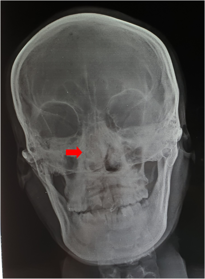
A 28-year-old man consulted an ear, nose, and throat (ENT) surgeon after complaints of headache for two days, a history of epistaxis, and vomitus. The right side of the nose had been punctured by wood as a result of falling from a motorcycle. A computed tomography (CT) scan led to diagnosis of a transnasal penetrating intracranial injury. Removal of the transcranial foreign body was carried out jointly by a neurosurgeon and ENT surgeon. Postoperatively, antibiotics were given for 14 days, and the patient was discharged without neurological deficit.
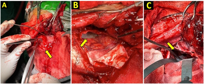
Early diagnostic procedures, such as CT scan of the skull to assess trajectory and extent of vascular and brain tissue injury, are required for appropriate surgical planning and post-operative treatment of such patients. Surgery was performed by combined transcranial and transnasal endoscopy to identify the skull base, dura mater defect, and brain tissue damage. Removal of the corpus alienum by transnasal endoscopy yielded a good outcome.
Combined transcranial and transnasal endoscopic approach showed better result than transcranial approach only. The wooden foreign body can be completely eliminated transnasally without active bleeding using this approach. The patient was discharged with good outcome.
经鼻穿透性颅脑损伤是一种罕见的创伤性脑损伤,可导致严重且致命的脑损伤以及高死亡率,需要立即进行多学科手术治疗。我们描述了一个不常见的病例,一名因意外脑部穿透伤就诊的患者被发现有一个经鼻穿透颅内的木质物体。
一名28岁男性因头痛两天、鼻出血史和呕吐前来咨询耳鼻喉科(ENT)外科医生。该患者因从摩托车上坠落,右侧鼻子被木头刺穿。计算机断层扫描(CT)检查诊断为经鼻穿透性颅脑损伤。神经外科医生和耳鼻喉科医生联合进行了经颅异物取出术。术后给予抗生素治疗14天,患者出院时无神经功能缺损。
对于此类患者,需要进行早期诊断程序,如头颅CT扫描以评估血管和脑组织损伤的轨迹及范围,以便进行适当的手术规划和术后治疗。通过经颅和经鼻内镜联合手术来确定颅底、硬脑膜缺损和脑组织损伤情况。经鼻内镜取出异物取得了良好效果。
经颅和经鼻内镜联合方法比单纯经颅方法效果更好。使用这种方法可以经鼻完全清除木质异物且无活动性出血。患者出院时情况良好。