Imaging Center, The First Affiliated Hospital of Xinjiang Medical University, No. 137, South Liyushan Road, Xinshi District, Urumqi, 830000, Xinjiang, China.
BMC Med Imaging. 2022 Aug 9;22(1):142. doi: 10.1186/s12880-022-00870-x.
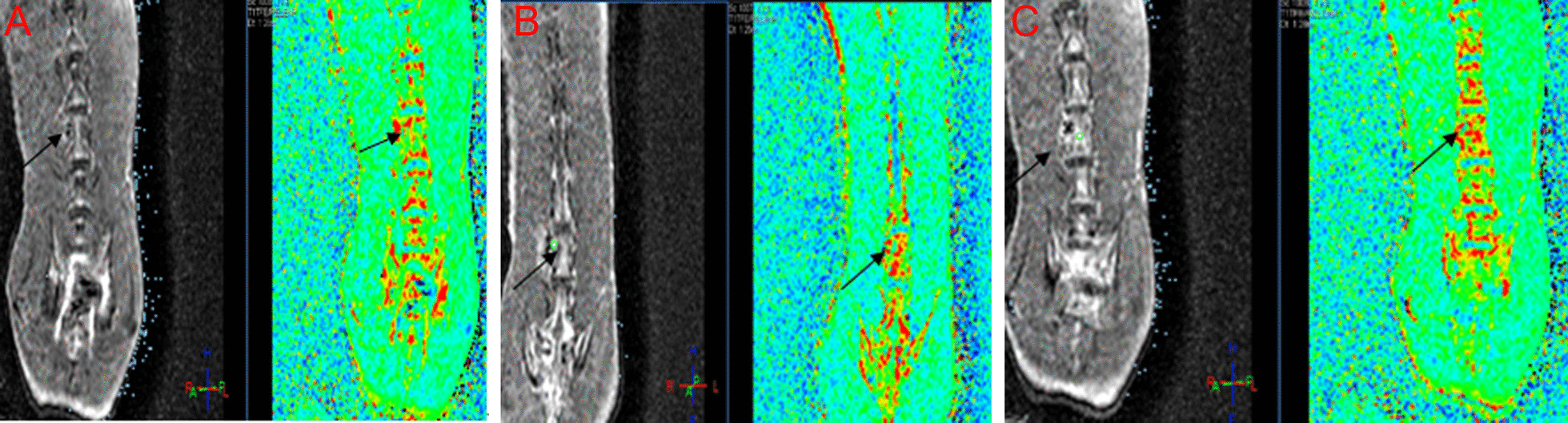
This study aimed to analyze the application value of magnetic resonance (MR)-perfusion weighted imaging (PWI) in the early imaging diagnosis of rabbit spinal tuberculosis.
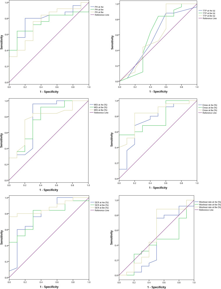
Spinal tuberculosis model was established using ATCC25177 Mycobacterium tuberculosis strain in the lumbar spine of rabbits. Forty rabbits were divided into 2 groups: rabbits in the experiment group were injected with 0.2 ml of 5.0 mg/ml tuberculosis suspension (n = 30) and those in the control group were injected with 0.2 ml of normal saline (n = 10) after vertebrae drilling surgery. Routine MRI and MR-PWI were performed at 4, 6, and 8 weeks after surgery. The statistical difference in terms of perfusion parameter values in the early MR-PWI scan of spinal tuberculosis between two groups was analyzed. The receiver operating characteristic (ROC) curve analysis was conducted for the accuracy of MR-PWI parameters in the early diagnosis of spinal tuberculosis.
Except time to peak, the other perfusion parameters in the experiment group were all increased with time. In addition, the difference between the two groups, as well as the differences at each time point was statistically significant (all P < 0.05). First-pass enhancement rate (Efirst), early enhancement rate (Ee), peak height (PH), maximum slope of increase (MSI), maximum signal enhancement rate (Emax) and signal enhancement rate (SER) showed high values in early diagnosing spinal tuberculosis.
The parameters including Efirst, Ee, PH, MSI, Emax and SER may provide valuable imaging evidence for the early diagnosis of spinal tuberculosis in clinical application.
本研究旨在分析磁共振(MR)灌注加权成像(PWI)在兔脊柱结核早期影像学诊断中的应用价值。
采用 ATCC25177 结核分枝杆菌株在兔腰椎建立脊柱结核模型。40 只兔随机分为实验组和对照组,每组 20 只。实验组经椎弓根钻孔术后注入 0.2ml 浓度为 5.0mg/ml 的结核混悬液(n=30),对照组注入 0.2ml 生理盐水(n=10)。术后 4、6、8 周分别行常规 MRI 和 MR-PWI 检查。分析两组兔脊柱结核早期 MR-PWI 扫描时灌注参数值的统计学差异。采用受试者工作特征(ROC)曲线分析 MR-PWI 参数对脊柱结核早期诊断的准确性。
实验组除达峰时间外,其余灌注参数均随时间增加而增加,且两组间差异及各时间点差异均有统计学意义(均 P<0.05)。首过增强率(Efirst)、早期增强率(Ee)、峰值高度(PH)、最大斜率(MSI)、最大信号增强率(Emax)和信号增强率(SER)在早期诊断脊柱结核时具有较高的诊断价值。
Efirst、Ee、PH、MSI、Emax 和 SER 等参数可能为临床应用中脊柱结核的早期诊断提供有价值的影像学依据。