Lineberger Comprehensive Cancer Center, The University of North Carolina at Chapel Hill, Chapel Hill, North Carolina, United States of America.
Division of Structural Biology, The Wellcome Trust Centre for Human Genetics, University of Oxford, Oxford, United Kingdom.
PLoS Pathog. 2022 Aug 15;18(8):e1010543. doi: 10.1371/journal.ppat.1010543. eCollection 2022 Aug.
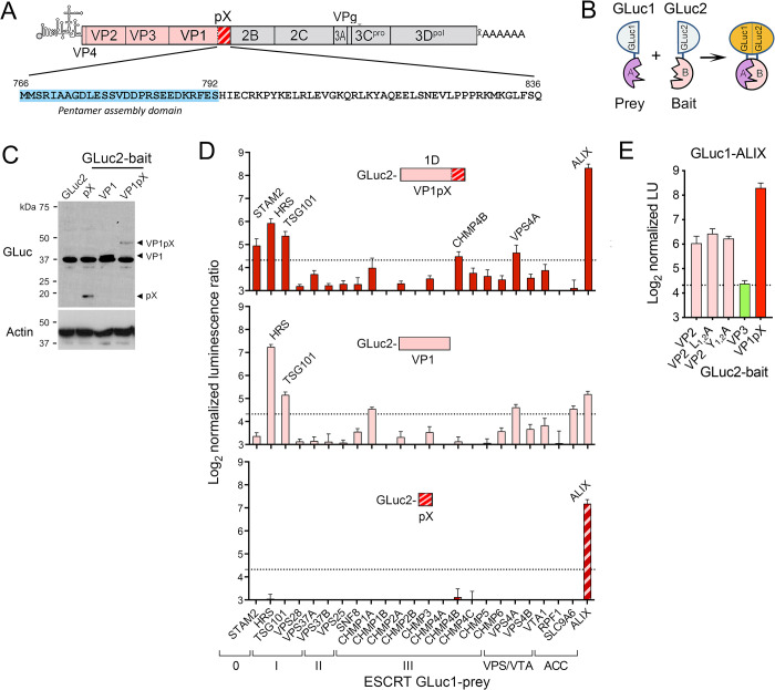
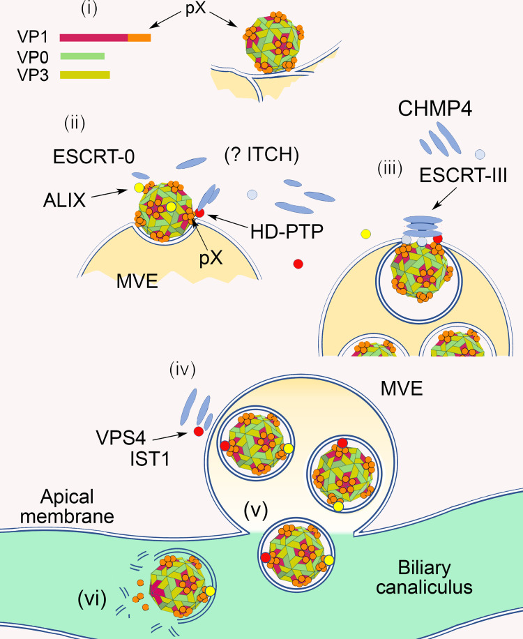
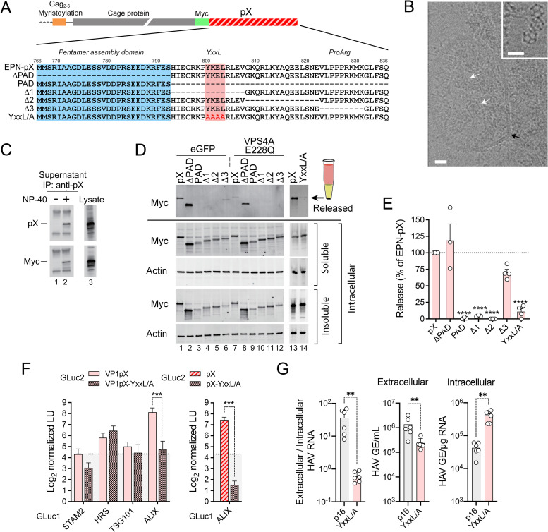
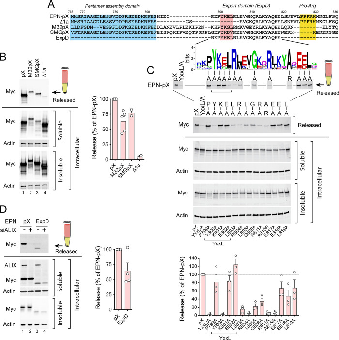
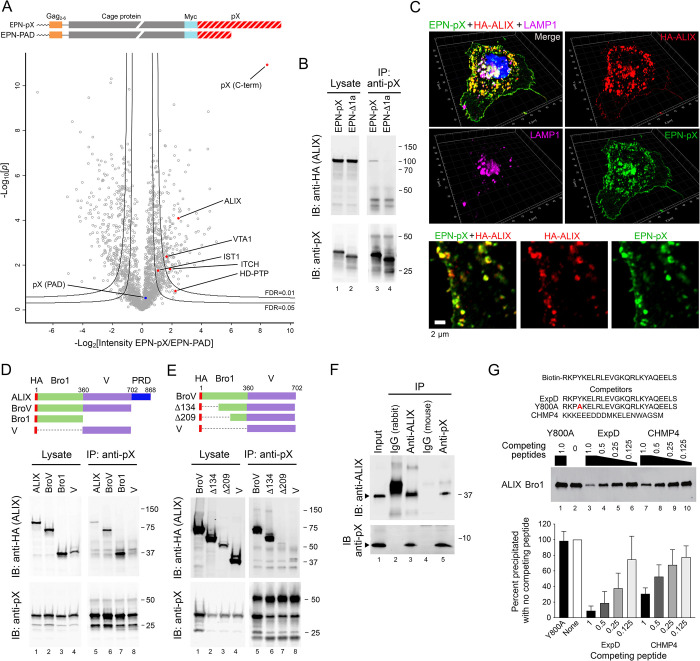
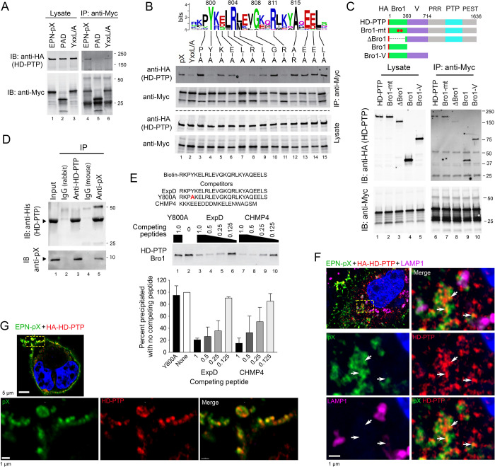
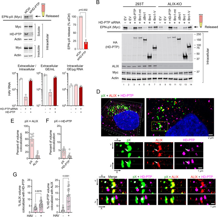
Although picornaviruses are conventionally considered 'nonenveloped', members of multiple picornaviral genera are released nonlytically from infected cells in extracellular vesicles. The mechanisms underlying this process are poorly understood. Here, we describe interactions of the hepatitis A virus (HAV) capsid with components of host endosomal sorting complexes required for transport (ESCRT) that play an essential role in release. We show release of quasi-enveloped virus (eHAV) in exosome-like vesicles requires a conserved export signal located within the 8 kDa C-terminal VP1 pX extension that functions in a manner analogous to late domains of canonical enveloped viruses. Fusing pX to a self-assembling engineered protein nanocage (EPN-pX) resulted in its ESCRT-dependent release in extracellular vesicles. Mutational analysis identified a 24 amino acid peptide sequence located within the center of pX that was both necessary and sufficient for nanocage release. Deleting a YxxL motif within this sequence ablated eHAV release, resulting in virus accumulating intracellularly. The pX export signal is conserved in non-human hepatoviruses from a wide range of mammalian species, and functional in pX sequences from bat hepatoviruses when fused to the nanocage protein, suggesting these viruses are released as quasi-enveloped virions. Quantitative proteomics identified multiple ESCRT-related proteins associating with EPN-pX, including ALG2-interacting protein X (ALIX), and its paralog, tyrosine-protein phosphatase non-receptor type 23 (HD-PTP), a second Bro1 domain protein linked to sorting of ubiquitylated cargo into multivesicular endosomes. RNAi-mediated depletion of either Bro1 domain protein impeded eHAV release. Super-resolution fluorescence microscopy demonstrated colocalization of viral capsids with endogenous ALIX and HD-PTP. Co-immunoprecipitation assays using biotin-tagged peptides and recombinant proteins revealed pX interacts directly through the export signal with N-terminal Bro1 domains of both HD-PTP and ALIX. Our study identifies an exceptionally potent viral export signal mediating extracellular release of virus-sized protein assemblies and shows release requires non-redundant activities of both HD-PTP and ALIX.
虽然小核糖核酸病毒通常被认为是“非包膜的”,但多个小核糖核酸病毒属的成员以细胞外囊泡的形式非裂解性地从感染细胞中释放。这一过程的机制还知之甚少。在这里,我们描述了甲型肝炎病毒(HAV)衣壳与参与运输所需的宿主内体分选复合物组成成分(ESCRT)之间的相互作用,该作用在释放中起着至关重要的作用。我们表明,类包膜病毒(eHAV)的释放需要位于 8 kDa C 末端 VP1 pX 延伸内的保守输出信号,该信号的作用方式类似于经典包膜病毒的晚期结构域。将 pX 融合到自组装的工程蛋白纳米笼(EPN-pX)中,导致其在细胞外囊泡中依赖 ESCRT 的释放。突变分析确定了 pX 中心的一个 24 个氨基酸肽序列,该序列既是必需的,也是充分的纳米笼释放。删除该序列中的 YxxL 基序会使 eHAV 释放失效,导致病毒在细胞内积累。该 pX 输出信号在来自广泛哺乳动物物种的非人类肝病毒中保守,并在与纳米笼蛋白融合的蝙蝠肝病毒的 pX 序列中具有功能,表明这些病毒以类包膜病毒的形式释放。定量蛋白质组学鉴定出与 EPN-pX 相关的多种 ESCRT 相关蛋白,包括 ALG2 相互作用蛋白 X(ALIX)及其同源物,酪氨酸蛋白磷酸酶非受体型 23(HD-PTP),这是一种与泛素化货物分选到多泡体中的第二个 Bro1 结构域蛋白。RNAi 介导的 Bro1 结构域蛋白耗竭会阻碍 eHAV 的释放。超分辨率荧光显微镜显示病毒衣壳与内源性 ALIX 和 HD-PTP 共定位。使用生物素标记的肽和重组蛋白进行的共免疫沉淀实验表明,pX 通过其输出信号与 HD-PTP 和 ALIX 的 N 末端 Bro1 结构域直接相互作用。我们的研究确定了一种非常有效的病毒输出信号,介导病毒大小的蛋白组装体的细胞外释放,并表明释放需要 HD-PTP 和 ALIX 的非冗余活性。